 Movie
Movie Controller


+ Open data

- Basic information
| Entry | Database: PDB / ID: 1f1o | ||||||
|---|---|---|---|---|---|---|---|
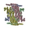
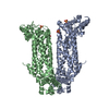
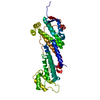
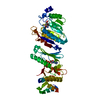
| Title | STRUCTURAL STUDIES OF ADENYLOSUCCINATE LYASES | ||||||
 Components Components | ADENYLOSUCCINATE LYASE | ||||||
 Keywords Keywords | LYASE / purine biosynthesis | ||||||
| Function / homology |  Function and homology information Function and homology informationAMP biosynthetic process / adenylosuccinate lyase / N6-(1,2-dicarboxyethyl)AMP AMP-lyase (fumarate-forming) activity / (S)-2-(5-amino-1-(5-phospho-D-ribosyl)imidazole-4-carboxamido) succinate lyase (fumarate-forming) activity / 'de novo' AMP biosynthetic process / 'de novo' IMP biosynthetic process / cytosolSimilarity search - Function | ||||||
| Biological species |  Bacillus subtilis (bacteria) Bacillus subtilis (bacteria) | ||||||
| Method | X-RAY DIFFRACTION / SYNCHROTRON / Resolution: 3.25 Å | ||||||
 Authors Authors | Toth, E.A. / Yeates, T. | ||||||
 Citation Citation | Journal: J.Mol.Biol. / Year: 2000 Title: The crystal structure of adenylosuccinate lyase from Pyrobaculum aerophilum reveals an intracellular protein with three disulfide bonds. Authors: Toth, E.A. / Worby, C. / Dixon, J.E. / Goedken, E.R. / Marqusee, S. / Yeates, T.O. | ||||||
| History |
| ||||||
| Remark 99 | The coordinate section in this entry contains CA atoms only |
- Structure visualization
| Structure viewer | Molecule: MolmilJmol/JSmol |
|---|
- Downloads & links
-Download
| PDBx/mmCIF format | 1f1o.cif.gz | 24.7 KB | Display | PDBx/mmCIF format |
|---|---|---|---|---|
| PDB format | pdb1f1o.ent.gz | 12.5 KB | Display | PDB format |
| PDBx/mmJSON format | 1f1o.json.gz | Tree view | PDBx/mmJSON format | |
| Others |  Other downloads Other downloads |
-Validation report
| Arichive directory | https://data.pdbj.org/pub/pdb/validation_reports/f1/1f1oftp://data.pdbj.org/pub/pdb/validation_reports/f1/1f1o | HTTPS FTP |
|---|
-Related structure data


-Links
 PDBj
PDBj

- Assembly
| Deposited unit | 
| ||||||||
|---|---|---|---|---|---|---|---|---|---|
| 1 |
| ||||||||
| Unit cell |
|
-Components
| #1: Protein | / ASL / Coordinate model: Cα atoms only Mass: 49553.734 Da / Num. of mol.: 1 Source method: isolated from a genetically manipulated source Source: (gene. exp.) Bacillus subtilis (bacteria) / Production host: Escherichia coli (E. coli) / References: UniProt: P12047, adenylosuccinate lyase |
|---|
-Experimental details
-Experiment
| Experiment | Method: X-RAY DIFFRACTION / Number of used crystals: 1 |
|---|
- Sample preparation
| Crystal | Density Matthews: 6.89 Å3/Da | ||||||||||||||||||||||||||||
|---|---|---|---|---|---|---|---|---|---|---|---|---|---|---|---|---|---|---|---|---|---|---|---|---|---|---|---|---|---|
| Crystal grow | Temperature: 293 K / Method: vapor diffusion, hanging drop / pH: 5.8 Details: 0.1 M MES, 1.5 M ammonium sulfate, pH 5.8, VAPOR DIFFUSION, HANGING DROP, temperature 293K | ||||||||||||||||||||||||||||
| Crystal | *PLUS Density % sol: 47.7 % | ||||||||||||||||||||||||||||
| Crystal grow | *PLUS Temperature: 18 ℃Details: This particular structure is not described in this paper. pH: 5 | ||||||||||||||||||||||||||||
| Components of the solutions | *PLUS
|
-Data collection
| Diffraction | Mean temperature: 100 K |
|---|---|
| Diffraction source | Source: SYNCHROTRON / Site: NSLS  / Beamline: X12C / Beamline: X12C |
| Detector | Type: MARRESEARCH / Detector: IMAGE PLATE |
| Radiation | Protocol: SINGLE WAVELENGTH / Monochromatic (M) / Laue (L): M / Scattering type: x-ray |
| Radiation wavelength | Relative weight: 1 |
| Reflection | Resolution: 3.25→50 Å / Num. all: 22850 / Num. obs: 2843 / % possible obs: 87.6 % / Observed criterion σ(I): -3 / Rmerge(I) obs: 0.058 |
| Reflection | *PLUS Num. measured all: 22850 |
- Processing
| Software |
| ||||||||||||||||||||
|---|---|---|---|---|---|---|---|---|---|---|---|---|---|---|---|---|---|---|---|---|---|
| Refinement | Resolution: 3.25→50 Å / Stereochemistry target values: Engh & Huber
| ||||||||||||||||||||
| Displacement parameters |
| ||||||||||||||||||||
| Refinement step | Cycle: LAST / Resolution: 3.25→50 Å
| ||||||||||||||||||||
| Refine LS restraints |
| ||||||||||||||||||||
| Xplor file | Serial no: 1 / Param file: PROTEIN_REP.PARAM | ||||||||||||||||||||
| Software | *PLUS Name: CNS / Classification: refinement | ||||||||||||||||||||
| Refinement | *PLUS Lowest resolution: 50 Å | ||||||||||||||||||||
| Solvent computation | *PLUS | ||||||||||||||||||||
| Displacement parameters | *PLUS |
