 Movie
Movie Controller

[English] 日本語
 Yorodumi
- PDB-4r9k: Structure of thermostable eightfold mutant of limonene epoxide hy... -
+ Open data

- Basic information
| Entry | Database: PDB / ID: 4r9k | ||||||
|---|---|---|---|---|---|---|---|
| Title | Structure of thermostable eightfold mutant of limonene epoxide hydrolase from Rhodococcus erythropolis | ||||||
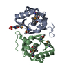
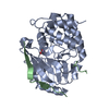
 Components Components | Limonene-1,2-epoxide hydrolase | ||||||
 Keywords Keywords | HYDROLASE / limonene epoxide hydrolase / NTF-2 fold / engineered / thermostable | ||||||
| Function / homology |  Function and homology informationlimonene-1,2-epoxide hydrolase / limonene-1,2-epoxide hydrolase activity Function and homology informationlimonene-1,2-epoxide hydrolase / limonene-1,2-epoxide hydrolase activitySimilarity search - Function | ||||||
| Biological species |  Rhodococcus erythropolis (bacteria) Rhodococcus erythropolis (bacteria) | ||||||
| Method | X-RAY DIFFRACTION / SYNCHROTRON / MOLECULAR REPLACEMENT / Resolution: 1.5 Å | ||||||
 Authors Authors | Floor, R.J. / Wijma, H.J. / Jekel, P.A. / Terwisscha van Scheltinga, A.C. / Dijkstra, B.W. / Janssen, D.B. | ||||||
 Citation Citation | Journal: Proteins / Year: 2015 Title: X-ray crystallographic validation of structure predictions used in computational design for protein stabilization. Authors: Floor, R.J. / Wijma, H.J. / Jekel, P.A. / Terwisscha van Scheltinga, A.C. / Dijkstra, B.W. / Janssen, D.B. | ||||||
| History |
|
- Structure visualization
| Structure viewer | Molecule: MolmilJmol/JSmol |
|---|
- Downloads & links
-Download
| PDBx/mmCIF format | 4r9k.cif.gz | 112 KB | Display | PDBx/mmCIF format |
|---|---|---|---|---|
| PDB format | pdb4r9k.ent.gz | 86.1 KB | Display | PDB format |
| PDBx/mmJSON format | 4r9k.json.gz | Tree view | PDBx/mmJSON format | |
| Others |  Other downloads Other downloads |
-Validation report
| Arichive directory | https://data.pdbj.org/pub/pdb/validation_reports/r9/4r9kftp://data.pdbj.org/pub/pdb/validation_reports/r9/4r9k | HTTPS FTP |
|---|
-Related structure data
| Related structure data |  4r9lC  1nwwS S: Starting model for refinement C: citing same article ( |
|---|---|
| Similar structure data |


-Links
 PDBj
PDBj
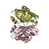
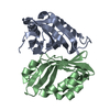
- Assembly
| Deposited unit | 
| ||||||||
|---|---|---|---|---|---|---|---|---|---|
| 1 | 
| ||||||||
| 2 | 
| ||||||||
| Unit cell |
| ||||||||
| Components on special symmetry positions |
|
-Components
| #1: Protein | Mass: 19482.072 Da / Num. of mol.: 3 / Mutation: S15P, A19K, E45K, T76K, T85V, N92K, Y96F, E124D Source method: isolated from a genetically manipulated source Source: (gene. exp.) Rhodococcus erythropolis (bacteria) / Gene: limA / Plasmid: pBAD / Production host: Escherichia coli (E. coli) / Strain (production host): TOP10 / References: UniProt: Q9ZAG3, limonene-1,2-epoxide hydrolase#2: Chemical | ChemComp-GOL / Glycerol  Mass: 92.094 Da / Num. of mol.: 5 / Source method: obtained synthetically / Formula: C3H8O3 Mass: 92.094 Da / Num. of mol.: 5 / Source method: obtained synthetically / Formula: C3H8O3#3: Chemical |   Mass: 131.173 Da / Num. of mol.: 3 / Source method: obtained synthetically / Formula: C6H13NO2 Mass: 131.173 Da / Num. of mol.: 3 / Source method: obtained synthetically / Formula: C6H13NO2#4: Water | ChemComp-HOH / | Water Mass: 18.015 Da / Num. of mol.: 362 / Source method: isolated from a natural source / Formula: H2O Mass: 18.015 Da / Num. of mol.: 362 / Source method: isolated from a natural source / Formula: H2O |
|---|
-Experimental details
-Experiment
| Experiment | Method: X-RAY DIFFRACTION / Number of used crystals: 1 |
|---|
- Sample preparation
| Crystal | Density Matthews: 2.11 Å3/Da / Density % sol: 41.72 % |
|---|---|
| Crystal grow | Temperature: 294 K / Method: vapor diffusion, sitting drop / pH: 6.4 Details: 1.44 M tri-sodium citrate, pH 6.4, VAPOR DIFFUSION, SITTING DROP, temperature 294K |
-Data collection
| Diffraction | Mean temperature: 77 K | |||||||||||||||||||||
|---|---|---|---|---|---|---|---|---|---|---|---|---|---|---|---|---|---|---|---|---|---|---|
| Diffraction source | Source: SYNCHROTRON / Site: ESRF  / Beamline: ID29 / Wavelength: 0.97625 Å / Beamline: ID29 / Wavelength: 0.97625 Å | |||||||||||||||||||||
| Detector | Type: DECTRIS PILATUS 6M / Detector: PIXEL / Date: Jul 21, 2012 | |||||||||||||||||||||
| Radiation | Monochromator: channel-cut Si(111) / Protocol: SINGLE WAVELENGTH / Monochromatic (M) / Laue (L): M / Scattering type: x-ray | |||||||||||||||||||||
| Radiation wavelength | Wavelength: 0.97625 Å / Relative weight: 1 | |||||||||||||||||||||
| Reflection | Resolution: 1.45→76.253 Å / Num. obs: 87794 / % possible obs: 99.9 % / Observed criterion σ(F): 2.9 / Observed criterion σ(I): 2.9 / Redundancy: 9.9 % / Biso Wilson estimate: 13.8 Å2 / Rmerge(I) obs: 0.073 / Rsym value: 0.024 / Net I/σ(I): 18 | |||||||||||||||||||||
| Reflection shell | Diffraction-ID: 1
|
- Processing
| Software |
| |||||||||||||||||||||||||||||||||||||||||||||||||||||||||||||||||||||||||||||||||||||||||||||||||||||||||
|---|---|---|---|---|---|---|---|---|---|---|---|---|---|---|---|---|---|---|---|---|---|---|---|---|---|---|---|---|---|---|---|---|---|---|---|---|---|---|---|---|---|---|---|---|---|---|---|---|---|---|---|---|---|---|---|---|---|---|---|---|---|---|---|---|---|---|---|---|---|---|---|---|---|---|---|---|---|---|---|---|---|---|---|---|---|---|---|---|---|---|---|---|---|---|---|---|---|---|---|---|---|---|---|---|---|---|
| Refinement | Method to determine structure: MOLECULAR REPLACEMENT Starting model: PDB ENTRY 1NWW Resolution: 1.5→44 Å / Cor.coef. Fo:Fc: 0.976 / Cor.coef. Fo:Fc free: 0.971 / SU B: 1.002 / SU ML: 0.038 / Cross valid method: THROUGHOUT / σ(F): 2 / ESU R: 0.056 / ESU R Free: 0.058 / Stereochemistry target values: MAXIMUM LIKELIHOOD / Details: HYDROGENS HAVE BEEN ADDED IN THE RIDING POSITIONS
| |||||||||||||||||||||||||||||||||||||||||||||||||||||||||||||||||||||||||||||||||||||||||||||||||||||||||
| Solvent computation | Ion probe radii: 0.8 Å / Shrinkage radii: 0.8 Å / VDW probe radii: 1.2 Å / Solvent model: MASK | |||||||||||||||||||||||||||||||||||||||||||||||||||||||||||||||||||||||||||||||||||||||||||||||||||||||||
| Displacement parameters | Biso mean: 18.991 Å2
| |||||||||||||||||||||||||||||||||||||||||||||||||||||||||||||||||||||||||||||||||||||||||||||||||||||||||
| Refinement step | Cycle: LAST / Resolution: 1.5→44 Å
| |||||||||||||||||||||||||||||||||||||||||||||||||||||||||||||||||||||||||||||||||||||||||||||||||||||||||
| Refine LS restraints |
| |||||||||||||||||||||||||||||||||||||||||||||||||||||||||||||||||||||||||||||||||||||||||||||||||||||||||
| LS refinement shell | Resolution: 1.5→1.539 Å / Total num. of bins used: 20
|
