 Movie
Movie Controller


+ Open data

- Basic information
| Entry | Database: PDB / ID: 3keh | |||||||||
|---|---|---|---|---|---|---|---|---|---|---|
| Title | Crystal Structure of N370S Glucocerebrosidase mutant at pH 7.4 | |||||||||
 Components Components | Glucocerebrosidase | |||||||||
 Keywords Keywords | HYDROLASE / Glucocerebrosidase / Acid-beta-Glucosidase / N370S / Glucosyceremidase / TIM Barrel / Alternative initiation / Disease mutation / Disulfide bond / Gaucher disease / Glycoprotein / Glycosidase / Ichthyosis / Lipid metabolism / Lysosome / Membrane / Sphingolipid metabolism | |||||||||
| Function / homology |  Function and homology information Function and homology informationpositive regulation of protein lipidation / steryl-beta-glucosidase activity / beta-glucoside catabolic process / positive regulation of neuronal action potential / cerebellar Purkinje cell layer formation / galactosylceramidase / termination of signal transduction / galactosylceramidase activity / positive regulation of autophagy of mitochondrion in response to mitochondrial depolarization / lymphocyte migration ...positive regulation of protein lipidation / steryl-beta-glucosidase activity / beta-glucoside catabolic process / positive regulation of neuronal action potential / cerebellar Purkinje cell layer formation / galactosylceramidase / termination of signal transduction / galactosylceramidase activity / positive regulation of autophagy of mitochondrion in response to mitochondrial depolarization / lymphocyte migration / glucosylceramidase / glucosylceramide catabolic process / scavenger receptor binding / regulation of lysosomal protein catabolic process / sphingosine biosynthetic process / autophagosome organization / glucosylceramidase activity / microglial cell proliferation / regulation of TOR signaling / glucosyltransferase activity / ceramide biosynthetic process / lipid storage / response to thyroid hormone / microglia differentiation / Glycosphingolipid catabolism / pyramidal neuron differentiation / lipid glycosylation / brain morphogenesis / response to pH / Hydrolases; Glycosylases; Glycosidases, i.e. enzymes that hydrolyse O- and S-glycosyl compounds / positive regulation of protein-containing complex disassembly / motor behavior / neuromuscular process / Transferases; Glycosyltransferases; Hexosyltransferases / hematopoietic stem cell proliferation / lysosome organization / response to testosterone / response to dexamethasone / Association of TriC/CCT with target proteins during biosynthesis / negative regulation of interleukin-6 production / homeostasis of number of cells / antigen processing and presentation / regulation of macroautophagy / establishment of skin barrier / negative regulation of protein-containing complex assembly / positive regulation of protein dephosphorylation / cell maturation / cholesterol metabolic process / respiratory electron transport chain / cellular response to starvation / lysosomal lumen / negative regulation of MAP kinase activity / determination of adult lifespan / trans-Golgi network / negative regulation of inflammatory response / autophagy / response to estrogen / positive regulation of proteasomal ubiquitin-dependent protein catabolic process / T cell differentiation in thymus / cellular response to tumor necrosis factor / proteasome-mediated ubiquitin-dependent protein catabolic process / neuron apoptotic process / negative regulation of neuron apoptotic process / lysosome / lysosomal membrane / signaling receptor binding / Golgi apparatus / endoplasmic reticulum / extracellular exosomeSimilarity search - Function | |||||||||
| Biological species |  Homo sapiens (human) Homo sapiens (human) | |||||||||
| Method | X-RAY DIFFRACTION / SYNCHROTRON / MOLECULAR REPLACEMENT / Resolution: 2.8 Å | |||||||||
 Authors Authors | Wei, R.R. / Boucher, S. / Pan, C.Q. / Edmunds, T. | |||||||||
 Citation Citation | Journal: To be Published Title: Crystal Structure of Glucocerebrosidase Containing the N370S mutation: Implication on Chaperon Therapy Authors: Wei, R.R. / Boucher, S. / Hughes, H. / Guziewica, N. / Vanpatten, S. / Pan, C.Q. / Edmunds, T. | |||||||||
| History |
|
- Structure visualization
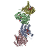
| Structure viewer | Molecule: MolmilJmol/JSmol |
|---|
- Downloads & links
-Download
| PDBx/mmCIF format | 3keh.cif.gz | 204.7 KB | Display | PDBx/mmCIF format |
|---|---|---|---|---|
| PDB format | pdb3keh.ent.gz | 164.4 KB | Display | PDB format |
| PDBx/mmJSON format | 3keh.json.gz | Tree view | PDBx/mmJSON format | |
| Others |  Other downloads Other downloads |
-Validation report
| Arichive directory | https://data.pdbj.org/pub/pdb/validation_reports/ke/3kehftp://data.pdbj.org/pub/pdb/validation_reports/ke/3keh | HTTPS FTP |
|---|
-Related structure data
| Related structure data | |
|---|---|
| Similar structure data |


-Links
 PDBj
PDBj


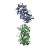
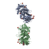
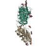
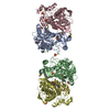
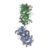
- Assembly
| Deposited unit | 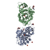
| ||||||||
|---|---|---|---|---|---|---|---|---|---|
| 1 |
| ||||||||
| 2 | 
| ||||||||
| 3 | 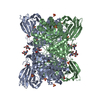
| ||||||||
| Unit cell |
|
-Components
-Protein , 1 types, 2 molecules AB
| #1: Protein | / Beta-glucocerebrosidase / Acid beta-glucosidase / D-glucosyl-N-acylsphingosine glucohydrolase / ...Beta-glucocerebrosidase / Acid beta-glucosidase / D-glucosyl-N-acylsphingosine glucohydrolase / Alglucerase / Imiglucerase Mass: 55632.195 Da / Num. of mol.: 2 / Mutation: N370S Source method: isolated from a genetically manipulated source Source: (gene. exp.) Homo sapiens (human) / Gene: GBA, GC, GLUC / Production host:   Spodoptera frugiperda (fall armyworm) / References: UniProt: P04062, glucosylceramidase Spodoptera frugiperda (fall armyworm) / References: UniProt: P04062, glucosylceramidase |
|---|
-Sugars , 3 types, 4 molecules 

| #2: Polysaccharide | 2-acetamido-2-deoxy-beta-D-glucopyranose-(1-4)-2-acetamido-2-deoxy-beta-D-glucopyranose / Mass: 424.401 Da / Num. of mol.: 1 Source method: isolated from a genetically manipulated source |
|---|---|
| #3: Sugar | ChemComp-NDG / N-Acetylglucosamine Type: D-saccharide, alpha linking / Mass: 221.208 Da / Num. of mol.: 1 Type: D-saccharide, alpha linking / Mass: 221.208 Da / Num. of mol.: 1Source method: isolated from a genetically manipulated source Formula: C8H15NO6 |
| #6: Sugar | N-Acetylglucosamine Type: D-saccharide, beta linking / Mass: 221.208 Da / Num. of mol.: 2 Type: D-saccharide, beta linking / Mass: 221.208 Da / Num. of mol.: 2Source method: isolated from a genetically manipulated source Formula: C8H15NO6 |
-Non-polymers , 3 types, 52 molecules 


| #4: Chemical | ChemComp-SO4 / Sulfate Mass: 96.063 Da / Num. of mol.: 8 / Source method: obtained synthetically / Formula: SO4 Mass: 96.063 Da / Num. of mol.: 8 / Source method: obtained synthetically / Formula: SO4#5: Chemical | Glycerol Mass: 92.094 Da / Num. of mol.: 2 / Source method: obtained synthetically / Formula: C3H8O3 Mass: 92.094 Da / Num. of mol.: 2 / Source method: obtained synthetically / Formula: C3H8O3#7: Water | ChemComp-HOH / | WaterMass: 18.015 Da / Num. of mol.: 42 / Source method: isolated from a natural source / Formula: H2O |
|---|
-Experimental details
-Experiment
| Experiment | Method: X-RAY DIFFRACTION / Number of used crystals: 1 |
|---|
- Sample preparation
| Crystal | Density Matthews: 3.19 Å3/Da / Density % sol: 61.4 % |
|---|---|
| Crystal grow | Temperature: 294 K / Method: vapor diffusion, sitting drop / pH: 7.4 Details: 0.7M Potassium Phosphate/0.7M Sodium Phosphate/0.1M HEPES, pH7.4, VAPOR DIFFUSION, SITTING DROP, temperature 294K |
-Data collection
| Diffraction | Mean temperature: 100 K |
|---|---|
| Diffraction source | Source: SYNCHROTRON / Site: ALS  / Beamline: 5.0.3 / Wavelength: 0.9765 Å / Beamline: 5.0.3 / Wavelength: 0.9765 Å |
| Detector | Type: ADSC QUANTUM 315r / Detector: CCD / Date: Feb 28, 2009 |
| Radiation | Monochromator: Single crystal, cylindrically bent, Si(220) / Protocol: SINGLE WAVELENGTH / Monochromatic (M) / Laue (L): M / Scattering type: x-ray |
| Radiation wavelength | Wavelength: 0.9765 Å / Relative weight: 1 |
| Reflection | Resolution: 2.8→85 Å / Num. all: 46396 / Num. obs: 31773 / % possible obs: 94.7 % / Observed criterion σ(F): 2 / Observed criterion σ(I): 2 / Redundancy: 5.2 % / Biso Wilson estimate: 39.5 Å2 / Rsym value: 0.158 / Net I/σ(I): 8.4 |
| Reflection shell | Resolution: 2.8→2.88 Å / Redundancy: 2.4 % / Mean I/σ(I) obs: 2.3 / Rsym value: 0.355 / % possible all: 91.1 |
- Processing
| Software |
| ||||||||||||||||||||||||||||||||||||||||||||||||||||||||||||||||||||||||||||||||||||||||||||||||||||||||||||||||||||||||||||||||||||||||||||||||||||||||||||||||||||||||||
|---|---|---|---|---|---|---|---|---|---|---|---|---|---|---|---|---|---|---|---|---|---|---|---|---|---|---|---|---|---|---|---|---|---|---|---|---|---|---|---|---|---|---|---|---|---|---|---|---|---|---|---|---|---|---|---|---|---|---|---|---|---|---|---|---|---|---|---|---|---|---|---|---|---|---|---|---|---|---|---|---|---|---|---|---|---|---|---|---|---|---|---|---|---|---|---|---|---|---|---|---|---|---|---|---|---|---|---|---|---|---|---|---|---|---|---|---|---|---|---|---|---|---|---|---|---|---|---|---|---|---|---|---|---|---|---|---|---|---|---|---|---|---|---|---|---|---|---|---|---|---|---|---|---|---|---|---|---|---|---|---|---|---|---|---|---|---|---|---|---|---|---|
| Refinement | Method to determine structure: MOLECULAR REPLACEMENT Starting model: !OGS Resolution: 2.8→50 Å / Cor.coef. Fo:Fc: 0.914 / Cor.coef. Fo:Fc free: 0.853 / SU B: 15.352 / SU ML: 0.303 / Cross valid method: THROUGHOUT / σ(F): 0 / σ(I): 0 / ESU R Free: 0.399 / Stereochemistry target values: MAXIMUM LIKELIHOOD / Details: HYDROGENS HAVE BEEN ADDED IN THE RIDING POSITIONS
| ||||||||||||||||||||||||||||||||||||||||||||||||||||||||||||||||||||||||||||||||||||||||||||||||||||||||||||||||||||||||||||||||||||||||||||||||||||||||||||||||||||||||||
| Solvent computation | Ion probe radii: 0.8 Å / Shrinkage radii: 0.8 Å / VDW probe radii: 1.4 Å / Solvent model: MASK | ||||||||||||||||||||||||||||||||||||||||||||||||||||||||||||||||||||||||||||||||||||||||||||||||||||||||||||||||||||||||||||||||||||||||||||||||||||||||||||||||||||||||||
| Displacement parameters | Biso mean: 28.113 Å2
| ||||||||||||||||||||||||||||||||||||||||||||||||||||||||||||||||||||||||||||||||||||||||||||||||||||||||||||||||||||||||||||||||||||||||||||||||||||||||||||||||||||||||||
| Refinement step | Cycle: LAST / Resolution: 2.8→50 Å
| ||||||||||||||||||||||||||||||||||||||||||||||||||||||||||||||||||||||||||||||||||||||||||||||||||||||||||||||||||||||||||||||||||||||||||||||||||||||||||||||||||||||||||
| Refine LS restraints |
| ||||||||||||||||||||||||||||||||||||||||||||||||||||||||||||||||||||||||||||||||||||||||||||||||||||||||||||||||||||||||||||||||||||||||||||||||||||||||||||||||||||||||||
| LS refinement shell | Resolution: 2.8→2.873 Å / Total num. of bins used: 20
|
