 Movie
Movie Controller


+ Open data

- Basic information
| Entry | Database: PDB / ID: 1ogs | |||||||||
|---|---|---|---|---|---|---|---|---|---|---|
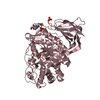
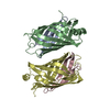
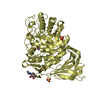
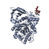
| Title | human acid-beta-glucosidase | |||||||||
 Components Components | Glucosylceramidase | |||||||||
 Keywords Keywords | HYDROLASE / GAUCHER DISEASE / GLUCOSIDASE / GLUCOCEREBROSIDASE / CEREZYME HYDROLASE / GLYCOSIDASE / SPHINGOLIPID METABOLISM / GLYCOPROTE LYSOSOME / MEMBRANE / DISEASE MUTATI POLYMORPHISM / ALTERNATIVE INITIATION / PHARMACEUTICAL / ISRAEL STRUCTURAL PROTEOMICS CENTER / ISPC / STRUCTURAL GENOMICS | |||||||||
| Function / homology |  Function and homology information Function and homology informationpositive regulation of protein lipidation / steryl-beta-glucosidase activity / beta-glucoside catabolic process / positive regulation of neuronal action potential / cerebellar Purkinje cell layer formation / galactosylceramidase / termination of signal transduction / galactosylceramidase activity / positive regulation of autophagy of mitochondrion in response to mitochondrial depolarization / lymphocyte migration ...positive regulation of protein lipidation / steryl-beta-glucosidase activity / beta-glucoside catabolic process / positive regulation of neuronal action potential / cerebellar Purkinje cell layer formation / galactosylceramidase / termination of signal transduction / galactosylceramidase activity / positive regulation of autophagy of mitochondrion in response to mitochondrial depolarization / lymphocyte migration / glucosylceramidase / glucosylceramide catabolic process / scavenger receptor binding / regulation of lysosomal protein catabolic process / sphingosine biosynthetic process / autophagosome organization / glucosylceramidase activity / microglial cell proliferation / regulation of TOR signaling / glucosyltransferase activity / ceramide biosynthetic process / lipid storage / response to thyroid hormone / microglia differentiation / Glycosphingolipid catabolism / pyramidal neuron differentiation / lipid glycosylation / brain morphogenesis / Hydrolases; Glycosylases; Glycosidases, i.e. enzymes that hydrolyse O- and S-glycosyl compounds / response to pH / positive regulation of protein-containing complex disassembly / motor behavior / neuromuscular process / Transferases; Glycosyltransferases; Hexosyltransferases / hematopoietic stem cell proliferation / lysosome organization / response to testosterone / response to dexamethasone / Association of TriC/CCT with target proteins during biosynthesis / negative regulation of interleukin-6 production / homeostasis of number of cells / antigen processing and presentation / regulation of macroautophagy / establishment of skin barrier / negative regulation of protein-containing complex assembly / positive regulation of protein dephosphorylation / cell maturation / respiratory electron transport chain / cellular response to starvation / cholesterol metabolic process / lysosomal lumen / negative regulation of MAP kinase activity / determination of adult lifespan / trans-Golgi network / autophagy / negative regulation of inflammatory response / response to estrogen / positive regulation of proteasomal ubiquitin-dependent protein catabolic process / T cell differentiation in thymus / cellular response to tumor necrosis factor / proteasome-mediated ubiquitin-dependent protein catabolic process / neuron apoptotic process / negative regulation of neuron apoptotic process / lysosome / lysosomal membrane / signaling receptor binding / Golgi apparatus / endoplasmic reticulum / extracellular exosomeSimilarity search - Function | |||||||||
| Biological species |  Homo sapiens (human) Homo sapiens (human) | |||||||||
| Method | X-RAY DIFFRACTION / SYNCHROTRON / MAD / Resolution: 2 Å | |||||||||
 Authors Authors | Dvir, H. / Harel, M. / McCarthy, A.A. / Toker, L. / Silman, I. / Futerman, A.H. / Sussman, J.L. | |||||||||
 Citation Citation | Journal: Embo Rep. / Year: 2003 Title: X-Ray Structure of Human Acid-Beta-Glucosidase, the Defective Enzyme in Gaucher Disease Authors: Dvir, H. / Harel, M. / Mccarthy, A.A. / Toker, L. / Silman, I. / Futerman, A.H. / Sussman, J.L. | |||||||||
| History |
| |||||||||
| Remark 700 | SHEET DETERMINATION METHOD: DSSP THE SHEETS PRESENTED AS "AC" IN EACH CHAIN ON SHEET RECORDS BELOW ... SHEET DETERMINATION METHOD: DSSP THE SHEETS PRESENTED AS "AC" IN EACH CHAIN ON SHEET RECORDS BELOW IS ACTUALLY AN 8-STRANDED BARREL THIS IS REPRESENTED BY A 9-STRANDED SHEET IN WHICH THE FIRST AND LAST STRANDS ARE IDENTICAL. THE SHEETS PRESENTED AS "BC" IN EACH CHAIN ON SHEET RECORDS BELOW IS ACTUALLY AN 8-STRANDED BARREL THIS IS REPRESENTED BY A 9-STRANDED SHEET IN WHICH THE FIRST AND LAST STRANDS ARE IDENTICAL. |
- Structure visualization
| Structure viewer | Molecule: MolmilJmol/JSmol |
|---|
- Downloads & links
-Download
| PDBx/mmCIF format | 1ogs.cif.gz | 225 KB | Display | PDBx/mmCIF format |
|---|---|---|---|---|
| PDB format | pdb1ogs.ent.gz | 184.5 KB | Display | PDB format |
| PDBx/mmJSON format | 1ogs.json.gz | Tree view | PDBx/mmJSON format | |
| Others |  Other downloads Other downloads |
-Validation report
| Arichive directory | https://data.pdbj.org/pub/pdb/validation_reports/og/1ogsftp://data.pdbj.org/pub/pdb/validation_reports/og/1ogs | HTTPS FTP |
|---|
-Related structure data
| Similar structure data |
|---|


-Links
 PDBj
PDBj


- Assembly
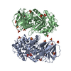
| Deposited unit | 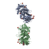
| |||||||||
|---|---|---|---|---|---|---|---|---|---|---|
| 1 | 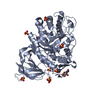
| |||||||||
| 2 | 
| |||||||||
| Unit cell |
| |||||||||
| Components on special symmetry positions |
|
-Components
| #1: Protein | / Acid beta-glucosidase / Alglucerase / Beta-glucocerebrosidase / Beta-GC / D-glucosyl-N- ...Acid beta-glucosidase / Alglucerase / Beta-glucocerebrosidase / Beta-GC / D-glucosyl-N-acylsphingosine glucohydrolase / Imiglucerase Mass: 55640.168 Da / Num. of mol.: 2 Source method: isolated from a genetically manipulated source Source: (gene. exp.) Homo sapiens (human) / Gene: GBA, GC, GLUC / Cell line (production host): CHO / Production host:   Cricetulus griseus (Chinese hamster) Cricetulus griseus (Chinese hamster)References: UniProt: P04062, glucosylceramidase, Transferases; Glycosyltransferases; Hexosyltransferases, steryl-beta-glucosidase#2: Polysaccharide | 2-acetamido-2-deoxy-beta-D-glucopyranose-(1-4)-2-acetamido-2-deoxy-beta-D-glucopyranose | / Mass: 424.401 Da / Num. of mol.: 1Source method: isolated from a genetically manipulated source #3: Chemical | ChemComp-SO4 / Sulfate  Mass: 96.063 Da / Num. of mol.: 15 / Source method: obtained synthetically / Formula: SO4 Mass: 96.063 Da / Num. of mol.: 15 / Source method: obtained synthetically / Formula: SO4#4: Sugar | ChemComp-NAG / | N-Acetylglucosamine  Type: D-saccharide, beta linking / Mass: 221.208 Da / Num. of mol.: 1 Type: D-saccharide, beta linking / Mass: 221.208 Da / Num. of mol.: 1Source method: isolated from a genetically manipulated source Formula: C8H15NO6 #5: Water | ChemComp-HOH / | Water Mass: 18.015 Da / Num. of mol.: 938 / Source method: isolated from a natural source / Formula: H2O Mass: 18.015 Da / Num. of mol.: 938 / Source method: isolated from a natural source / Formula: H2O |
|---|
-Experimental details
-Experiment
| Experiment | Method: X-RAY DIFFRACTION / Number of used crystals: 2 |
|---|
- Sample preparation
| Crystal | Density Matthews: 2.9 Å3/Da / Density % sol: 57.5 % | |||||||||||||||||||||||||||||||||||||||||||||||||||||||||||||||
|---|---|---|---|---|---|---|---|---|---|---|---|---|---|---|---|---|---|---|---|---|---|---|---|---|---|---|---|---|---|---|---|---|---|---|---|---|---|---|---|---|---|---|---|---|---|---|---|---|---|---|---|---|---|---|---|---|---|---|---|---|---|---|---|---|
| Crystal grow | Method: vapor diffusion, hanging drop / pH: 4.6 Details: VAPOUR DIFFUSION AT ROOM TEMP DROP CONTAINS 1.5 MICROLITER OF PROTEIN SOLUTION (10MG/ML) AND 1.5 MICROLITER OF MOTHER LIQUOR. MOTHER LIQUOR CONTAINS 1M AMMONIUM SULFATE, 0.17M GUANIDINE ...Details: VAPOUR DIFFUSION AT ROOM TEMP DROP CONTAINS 1.5 MICROLITER OF PROTEIN SOLUTION (10MG/ML) AND 1.5 MICROLITER OF MOTHER LIQUOR. MOTHER LIQUOR CONTAINS 1M AMMONIUM SULFATE, 0.17M GUANIDINE HYDROCHLORIDE, 0.02M KCL, 0.1M ACETATE PH 4.6. | |||||||||||||||||||||||||||||||||||||||||||||||||||||||||||||||
| Crystal grow | *PLUS Temperature: 19 ℃ / pH: 6.6 / Method: vapor diffusion, hanging drop | |||||||||||||||||||||||||||||||||||||||||||||||||||||||||||||||
| Components of the solutions | *PLUS
|
-Data collection
| Diffraction | Mean temperature: 100 K | |||||||||||||||
|---|---|---|---|---|---|---|---|---|---|---|---|---|---|---|---|---|
| Diffraction source | Source: SYNCHROTRON / Site: ESRF  / Beamline: ID14-4 / Wavelength: 1.0092, 1.0075, 1.0015, 0.8856 / Beamline: ID14-4 / Wavelength: 1.0092, 1.0075, 1.0015, 0.8856 | |||||||||||||||
| Detector | Date: Mar 15, 2003 | |||||||||||||||
| Radiation | Protocol: MAD / Monochromatic (M) / Laue (L): M / Scattering type: x-ray | |||||||||||||||
| Radiation wavelength |
| |||||||||||||||
| Reflection | Resolution: 2→10 Å / Num. obs: 93248 / % possible obs: 98.4 % / Redundancy: 10 % / Biso Wilson estimate: 15.6 Å2 / Rmerge(I) obs: 0.065 / Net I/σ(I): 21.6 | |||||||||||||||
| Reflection shell | Resolution: 2→2.1 Å / Redundancy: 3.5 % / Rmerge(I) obs: 0.37 / Mean I/σ(I) obs: 1.23 / % possible all: 84.7 | |||||||||||||||
| Reflection | *PLUS Highest resolution: 2 Å / Lowest resolution: 14.4 Å / Rmerge(I) obs: 0.084 | |||||||||||||||
| Reflection shell | *PLUS % possible obs: 98.3 % / Rmerge(I) obs: 0.373 / Mean I/σ(I) obs: 1.6 |
- Processing
| Software |
| ||||||||||||||||||||||||||||||||||||||||||||||||||||||||||||||||||||||||||||||||
|---|---|---|---|---|---|---|---|---|---|---|---|---|---|---|---|---|---|---|---|---|---|---|---|---|---|---|---|---|---|---|---|---|---|---|---|---|---|---|---|---|---|---|---|---|---|---|---|---|---|---|---|---|---|---|---|---|---|---|---|---|---|---|---|---|---|---|---|---|---|---|---|---|---|---|---|---|---|---|---|---|---|
| Refinement | Method to determine structure: MAD / Resolution: 2→14.4 Å / Rfactor Rfree error: 0.003 / Data cutoff high absF: 10000 / Isotropic thermal model: RESTRAINED / Cross valid method: THROUGHOUT / σ(F): 0
| ||||||||||||||||||||||||||||||||||||||||||||||||||||||||||||||||||||||||||||||||
| Solvent computation | Solvent model: FLAT MODEL / Bsol: 69.0691 Å2 / ksol: 0.391893 e/Å3 | ||||||||||||||||||||||||||||||||||||||||||||||||||||||||||||||||||||||||||||||||
| Displacement parameters | Biso mean: 28.7 Å2
| ||||||||||||||||||||||||||||||||||||||||||||||||||||||||||||||||||||||||||||||||
| Refine analyze |
| ||||||||||||||||||||||||||||||||||||||||||||||||||||||||||||||||||||||||||||||||
| Refinement step | Cycle: LAST / Resolution: 2→14.4 Å
| ||||||||||||||||||||||||||||||||||||||||||||||||||||||||||||||||||||||||||||||||
| Refine LS restraints |
| ||||||||||||||||||||||||||||||||||||||||||||||||||||||||||||||||||||||||||||||||
| LS refinement shell | Resolution: 2→2.13 Å / Rfactor Rfree error: 0.012 / Total num. of bins used: 6
| ||||||||||||||||||||||||||||||||||||||||||||||||||||||||||||||||||||||||||||||||
| Xplor file |
| ||||||||||||||||||||||||||||||||||||||||||||||||||||||||||||||||||||||||||||||||
| Refinement | *PLUS Highest resolution: 2 Å | ||||||||||||||||||||||||||||||||||||||||||||||||||||||||||||||||||||||||||||||||
| Solvent computation | *PLUS | ||||||||||||||||||||||||||||||||||||||||||||||||||||||||||||||||||||||||||||||||
| Displacement parameters | *PLUS | ||||||||||||||||||||||||||||||||||||||||||||||||||||||||||||||||||||||||||||||||
| Refine LS restraints | *PLUS
|
