 Movie
Movie Controller

[English] 日本語
 Yorodumi
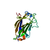
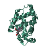
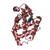
- PDB-3k7j: Crystal structure of the D100E mutant of the Indian Hedgehog N-te... -
+ Open data

- Basic information
| Entry | Database: PDB / ID: 3k7j | ||||||
|---|---|---|---|---|---|---|---|
| Title | Crystal structure of the D100E mutant of the Indian Hedgehog N-terminal signalling domain | ||||||
 Components Components | Indian hedgehog protein | ||||||
 Keywords Keywords | SIGNALING PROTEIN / alpha+beta sandwich / Autocatalytic cleavage / Cell membrane / Developmental protein / Disease mutation / Glycoprotein / Hydrolase / Lipoprotein / Membrane / Palmitate / Protease / Secreted | ||||||
| Function / homology |  Function and homology information Function and homology informationvitelline membrane formation / negative regulation of eye pigmentation / camera-type eye photoreceptor cell fate commitment / chondrocyte differentiation involved in endochondral bone morphogenesis / embryonic skeletal joint development / Formation of lateral plate mesoderm / negative regulation of alpha-beta T cell differentiation / embryonic camera-type eye morphogenesis / cholesterol-protein transferase activity / HHAT G278V doesn't palmitoylate Hh-Np ...vitelline membrane formation / negative regulation of eye pigmentation / camera-type eye photoreceptor cell fate commitment / chondrocyte differentiation involved in endochondral bone morphogenesis / embryonic skeletal joint development / Formation of lateral plate mesoderm / negative regulation of alpha-beta T cell differentiation / embryonic camera-type eye morphogenesis / cholesterol-protein transferase activity / HHAT G278V doesn't palmitoylate Hh-Np / RUNX2 regulates chondrocyte maturation / Ligand-receptor interactions / proteoglycan metabolic process / negative regulation of immature T cell proliferation in thymus / chondrocyte proliferation / negative regulation of T cell differentiation in thymus / retinal pigment epithelium development / positive regulation of T cell differentiation in thymus / epithelial cell-cell adhesion / Activation of SMO / patched binding / embryonic digestive tract morphogenesis / somite development / smooth muscle tissue development / epithelial cell morphogenesis / self proteolysis / head morphogenesis / embryonic pattern specification / Release of Hh-Np from the secreting cell / intein-mediated protein splicing / positive regulation of smoothened signaling pathway / pancreas development / negative regulation of chondrocyte differentiation / Class B/2 (Secretin family receptors) / regulation of growth / cell fate specification / positive regulation of alpha-beta T cell differentiation / positive regulation of mesenchymal cell proliferation / smoothened signaling pathway / branching involved in blood vessel morphogenesis / embryonic digit morphogenesis / heart looping / protein autoprocessing / positive regulation of collagen biosynthetic process / maternal process involved in female pregnancy / neuron development / response to mechanical stimulus / cell maturation / bone resorption / extracellular matrix / skeletal system development / positive regulation of epithelial cell proliferation / liver regeneration / Hedgehog ligand biogenesis / Hedgehog 'on' state / multicellular organism growth / osteoblast differentiation / cell-cell signaling / response to estradiol / peptidase activity / regulation of gene expression / in utero embryonic development / Hydrolases; Acting on ester bonds / Golgi membrane / calcium ion binding / endoplasmic reticulum membrane / negative regulation of apoptotic process / positive regulation of transcription by RNA polymerase II / extracellular space / plasma membraneSimilarity search - Function | ||||||
| Biological species |  Homo sapiens (human) Homo sapiens (human) | ||||||
| Method | X-RAY DIFFRACTION / MOLECULAR REPLACEMENT / Resolution: 1.9 Å | ||||||
 Authors Authors | He, Y.-X. / Kang, Y. / Zhang, W.J. / Yu, J. / Ma, G. / Zhou, C.-Z. | ||||||
 Citation Citation | Journal: To be Published Title: Crystal structure of the D100E mutant of the Indian Hedgehog N-terminal signalling domain Authors: He, Y.-X. / Kang, Y. / Zhang, W.J. / Yu, J. / Ma, G. / Zhou, C.-Z. | ||||||
| History |
|
- Structure visualization
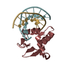
| Structure viewer | Molecule: MolmilJmol/JSmol |
|---|
- Downloads & links
-Download
| PDBx/mmCIF format | 3k7j.cif.gz | 47.9 KB | Display | PDBx/mmCIF format |
|---|---|---|---|---|
| PDB format | pdb3k7j.ent.gz | 31.7 KB | Display | PDB format |
| PDBx/mmJSON format | 3k7j.json.gz | Tree view | PDBx/mmJSON format | |
| Others |  Other downloads Other downloads |
-Validation report
| Arichive directory | https://data.pdbj.org/pub/pdb/validation_reports/k7/3k7jftp://data.pdbj.org/pub/pdb/validation_reports/k7/3k7j | HTTPS FTP |
|---|
-Related structure data
| Related structure data |  1vhhS S: Starting model for refinement |
|---|---|
| Similar structure data |


-Links
 PDBj
PDBj


- Assembly
| Deposited unit | 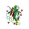
| ||||||||
|---|---|---|---|---|---|---|---|---|---|
| 1 |
| ||||||||
| Unit cell |
|
-Components
| #1: Protein | / IHH / HHG-2 Mass: 21129.658 Da / Num. of mol.: 1 / Fragment: Indian hedgehog protein N-product / Mutation: D100E Source method: isolated from a genetically manipulated source Source: (gene. exp.) Homo sapiens (human) / Gene: IHH / Plasmid: pET28a / Production host:  Escherichia coli (E. coli) / Strain (production host): Rosseta / References: UniProt: Q14623 Escherichia coli (E. coli) / Strain (production host): Rosseta / References: UniProt: Q14623 | ||||
|---|---|---|---|---|---|
| #2: Chemical | ChemComp-ZN /   Mass: 65.409 Da / Num. of mol.: 1 / Source method: obtained synthetically / Formula: Zn Mass: 65.409 Da / Num. of mol.: 1 / Source method: obtained synthetically / Formula: Zn | ||||
| #3: Chemical | ChemComp-SO4 / Sulfate  Mass: 96.063 Da / Num. of mol.: 5 / Source method: obtained synthetically / Formula: SO4 Mass: 96.063 Da / Num. of mol.: 5 / Source method: obtained synthetically / Formula: SO4#4: Chemical | ChemComp-CO3 / | Carbonate  Mass: 60.009 Da / Num. of mol.: 1 / Source method: obtained synthetically / Formula: CO3 Mass: 60.009 Da / Num. of mol.: 1 / Source method: obtained synthetically / Formula: CO3#5: Water | ChemComp-HOH / | Water Mass: 18.015 Da / Num. of mol.: 102 / Source method: isolated from a natural source / Formula: H2O Mass: 18.015 Da / Num. of mol.: 102 / Source method: isolated from a natural source / Formula: H2O |
-Experimental details
-Experiment
| Experiment | Method: X-RAY DIFFRACTION / Number of used crystals: 1 |
|---|
- Sample preparation
| Crystal | Density Matthews: 1.81 Å3/Da / Density % sol: 31.99 % |
|---|---|
| Crystal grow | Temperature: 289 K / Method: vapor diffusion, sitting drop / pH: 8 Details: 30% PEG 4000, 0.2M (NH4)2SO4, pH 8.0, VAPOR DIFFUSION, SITTING DROP, temperature 289K |
-Data collection
| Diffraction | Mean temperature: 100 K |
|---|---|
| Diffraction source | Source: ROTATING ANODE / Type: RIGAKU MICROMAX-007 HF / Wavelength: 1.54178 Å |
| Detector | Type: MAR scanner 345 mm plate / Detector: IMAGE PLATE / Date: Feb 8, 2009 |
| Radiation | Protocol: SINGLE WAVELENGTH / Monochromatic (M) / Laue (L): M / Scattering type: x-ray |
| Radiation wavelength | Wavelength: 1.54178 Å / Relative weight: 1 |
| Reflection | Resolution: 1.9→24.73 Å / Num. obs: 11849 / % possible obs: 98.8 % / Redundancy: 3.1 % / Biso Wilson estimate: 23.67 Å2 / Rmerge(I) obs: 0.079 / Net I/σ(I): 10.7 |
| Reflection shell | Resolution: 1.9→2 Å / Redundancy: 3.1 % / Rmerge(I) obs: 0.079 / Mean I/σ(I) obs: 2.4 / Num. unique all: 1680 / % possible all: 97.5 |
- Processing
| Software |
| |||||||||||||||||||||||||||||||||||
|---|---|---|---|---|---|---|---|---|---|---|---|---|---|---|---|---|---|---|---|---|---|---|---|---|---|---|---|---|---|---|---|---|---|---|---|---|
| Refinement | Method to determine structure: MOLECULAR REPLACEMENT Starting model: PDB ENTRY 1VHH Resolution: 1.9→18.721 Å / Occupancy max: 1 / Occupancy min: 0.49 / FOM work R set: 0.808 / SU ML: 0.24 / Cross valid method: THROUGHOUT / σ(F): 1.35 / Phase error: 26.46 / Stereochemistry target values: ML
| |||||||||||||||||||||||||||||||||||
| Solvent computation | Shrinkage radii: 0.9 Å / VDW probe radii: 1.11 Å / Solvent model: FLAT BULK SOLVENT MODEL / Bsol: 50.547 Å2 / ksol: 0.443 e/Å3 | |||||||||||||||||||||||||||||||||||
| Displacement parameters | Biso max: 59.32 Å2 / Biso mean: 26.239 Å2 / Biso min: 14.64 Å2
| |||||||||||||||||||||||||||||||||||
| Refinement step | Cycle: LAST / Resolution: 1.9→18.721 Å
| |||||||||||||||||||||||||||||||||||
| Refine LS restraints |
| |||||||||||||||||||||||||||||||||||
| LS refinement shell |
|
