 Movie
Movie Controller

[English] 日本語
 Yorodumi
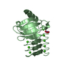
- PDB-4yeo: Triclinic HEWL co-crystallised with cisplatin, studied at a data ... -
+ Open data

- Basic information
| Entry | Database: PDB / ID: 4yeo | ||||||
|---|---|---|---|---|---|---|---|
| Title | Triclinic HEWL co-crystallised with cisplatin, studied at a data collection temperature of 150K - new refinement | ||||||
 Components Components | Lysozyme C | ||||||
 Keywords Keywords |  HYDROLASE / Re-refinement of 4mwk / lysozyme / metal binding HYDROLASE / Re-refinement of 4mwk / lysozyme / metal binding | ||||||
| Function / homology |  Function and homology information Function and homology informationLactose synthesis / Antimicrobial peptides / Neutrophil degranulation / beta-N-acetylglucosaminidase activity / cell wall macromolecule catabolic process / lysozyme / lysozyme activity / killing of cells of another organism / defense response to Gram-negative bacterium / defense response to Gram-positive bacterium ...Lactose synthesis / Antimicrobial peptides / Neutrophil degranulation / beta-N-acetylglucosaminidase activity / cell wall macromolecule catabolic process / lysozyme / lysozyme activity / killing of cells of another organism / defense response to Gram-negative bacterium / defense response to Gram-positive bacterium / defense response to bacterium / endoplasmic reticulum / extracellular space / identical protein binding / cytoplasmSimilarity search - Function | ||||||
| Biological species |  Gallus gallus (chicken) Gallus gallus (chicken) | ||||||
| Method | X-RAY DIFFRACTION / FOURIER SYNTHESIS / Resolution: 0.98 Å | ||||||
 Authors Authors | Shabalin, I.G. / Dauter, Z. / Jaskolski, M. / Minor, W. / Wlodawer, A. | ||||||
| Funding support |  United States, 1items United States, 1items
| ||||||
 Citation Citation | Journal: Acta Crystallogr.,Sect.D / Year: 2015 Title: Crystallography and chemistry should always go together: a cautionary tale of protein complexes with cisplatin and carboplatin. Authors: Shabalin, I. / Dauter, Z. / Jaskolski, M. / Minor, W. / Wlodawer, A. #1: Journal: Struct Dyn / Year: 2014 Title: Structural dynamics of cisplatin binding to histidine in a protein. Authors: Tanley, S.W. / Helliwell, J.R. | ||||||
| History |
|
- Structure visualization
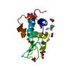
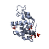
| Structure viewer | Molecule: MolmilJmol/JSmol |
|---|
- Downloads & links
-Download
| PDBx/mmCIF format | 4yeo.cif.gz | 80.7 KB | Display | PDBx/mmCIF format |
|---|---|---|---|---|
| PDB format | pdb4yeo.ent.gz | 57.8 KB | Display | PDB format |
| PDBx/mmJSON format | 4yeo.json.gz | Tree view | PDBx/mmJSON format | |
| Others |  Other downloads Other downloads |
-Validation report
| Arichive directory | https://data.pdbj.org/pub/pdb/validation_reports/ye/4yeoftp://data.pdbj.org/pub/pdb/validation_reports/ye/4yeo | HTTPS FTP |
|---|
-Related structure data
| Related structure data | 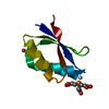 4ydxC  4yeaC  4yemC 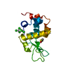 4yenC  4mwk S: Starting model for refinement C: citing same article ( |
|---|---|
| Similar structure data |


-Links
 PDBj
PDBj


- Assembly
| Deposited unit | 
| ||||||||
|---|---|---|---|---|---|---|---|---|---|
| 1 |
| ||||||||
| Unit cell |
|
-Components
-Protein , 1 types, 1 molecules A
| #1: Protein | Mass: 14289.080 Da / Num. of mol.: 1 / Source method: isolated from a natural source / Source: (natural) Gallus gallus (chicken) / References: UniProt: P00698, lysozyme |
|---|
-Non-polymers , 7 types, 232 molecules 


| #2: Chemical | ChemComp-CPT / Cisplatin Mass: 300.045 Da / Num. of mol.: 1 / Source method: obtained synthetically / Formula: Cl2H6N2Pt / Comment: medication, chemotherapy*YM Mass: 300.045 Da / Num. of mol.: 1 / Source method: obtained synthetically / Formula: Cl2H6N2Pt / Comment: medication, chemotherapy*YM | ||||||||||
|---|---|---|---|---|---|---|---|---|---|---|---|
| #3: Chemical | ChemComp-PT /  Mass: 195.078 Da / Num. of mol.: 4 / Source method: obtained synthetically / Formula: Pt Mass: 195.078 Da / Num. of mol.: 4 / Source method: obtained synthetically / Formula: Pt#4: Chemical | ChemComp-EDO / | Ethylene glycol Mass: 62.068 Da / Num. of mol.: 1 / Source method: obtained synthetically / Formula: C2H6O2 Mass: 62.068 Da / Num. of mol.: 1 / Source method: obtained synthetically / Formula: C2H6O2#5: Chemical | Dimethyl sulfoxide Mass: 78.133 Da / Num. of mol.: 2 / Source method: obtained synthetically / Formula: C2H6OS / Comment: DMSO, precipitant*YM Mass: 78.133 Da / Num. of mol.: 2 / Source method: obtained synthetically / Formula: C2H6OS / Comment: DMSO, precipitant*YM#6: Chemical | ChemComp-NO3 / Nitrate Mass: 62.005 Da / Num. of mol.: 7 / Source method: obtained synthetically / Formula: NO3 Mass: 62.005 Da / Num. of mol.: 7 / Source method: obtained synthetically / Formula: NO3#7: Chemical | ChemComp-ACT / | Acetate Mass: 59.044 Da / Num. of mol.: 1 / Source method: obtained synthetically / Formula: C2H3O2 Mass: 59.044 Da / Num. of mol.: 1 / Source method: obtained synthetically / Formula: C2H3O2#8: Water | ChemComp-HOH / | WaterMass: 18.015 Da / Num. of mol.: 216 / Source method: isolated from a natural source / Formula: H2O |
-Experimental details
-Experiment
| Experiment | Method: X-RAY DIFFRACTION / Number of used crystals: 1 |
|---|
- Sample preparation
| Crystal | Density Matthews: 1.89 Å3/Da / Density % sol: 31.34 % |
|---|---|
| Crystal grow | Temperature: 294 K / Method: evaporation / pH: 4.7 Details: 40mg HEWL co-crystallised with 2.6mg cisplatin, with the platinum compounds being in a 3-fold molar excess to the protein. 462.5 ul of a 0.02M NaAc solution along with 462.5 ul of a 0.5M ...Details: 40mg HEWL co-crystallised with 2.6mg cisplatin, with the platinum compounds being in a 3-fold molar excess to the protein. 462.5 ul of a 0.02M NaAc solution along with 462.5 ul of a 0.5M NaNo3 solution was used with 75 ul DMSO added |
-Data collection
| Diffraction | Mean temperature: 150 K |
|---|---|
| Diffraction source | Source: ROTATING ANODE / Type: BRUKER AXS MICROSTAR / Wavelength: 1.5418 Å |
| Detector | Type: APEX II CCD / Detector: CCD / Date: Dec 7, 2012 |
| Radiation | Monochromator: CONFOCAL MIRROR OPTICS / Protocol: SINGLE WAVELENGTH / Monochromatic (M) / Laue (L): M / Scattering type: x-ray |
| Radiation wavelength | Wavelength: 1.5418 Å / Relative weight: 1 |
| Reflection | Resolution: 0.98→29.28 Å / Num. obs: 48959 / % possible obs: 94.8 % / Observed criterion σ(I): 2 / Redundancy: 3.5 % / Biso Wilson estimate: 6 Å2 / Rmerge(I) obs: 0.045 / Rsym value: 0.045 / Net I/σ(I): 15.6 |
| Reflection shell | Resolution: 0.98→1.02 Å / Redundancy: 0.6 % / Rmerge(I) obs: 0.209 / Mean I/σ(I) obs: 3.2 / % possible all: 51.7 |
- Processing
| Software |
| ||||||||||||||||||||||||||||||||||||||||||||||||||||||||||||||||||||||||||||||||||||||||||
|---|---|---|---|---|---|---|---|---|---|---|---|---|---|---|---|---|---|---|---|---|---|---|---|---|---|---|---|---|---|---|---|---|---|---|---|---|---|---|---|---|---|---|---|---|---|---|---|---|---|---|---|---|---|---|---|---|---|---|---|---|---|---|---|---|---|---|---|---|---|---|---|---|---|---|---|---|---|---|---|---|---|---|---|---|---|---|---|---|---|---|---|
| Refinement | Method to determine structure: FOURIER SYNTHESIS Starting model: 4mwk 4mwk Resolution: 0.98→29.26 Å / Cor.coef. Fo:Fc: 0.982 / Cor.coef. Fo:Fc free: 0.977 / WRfactor Rfree: 0.139 / WRfactor Rwork: 0.119 / FOM work R set: 0.9356 / SU B: 0.558 / SU ML: 0.014 / SU R Cruickshank DPI: 0.0219 / SU Rfree: 0.0223 / Cross valid method: THROUGHOUT / σ(F): 0 / ESU R: 0.022 / ESU R Free: 0.022 / Stereochemistry target values: MAXIMUM LIKELIHOOD Details: HYDROGENS HAVE BEEN ADDED IN THE RIDING POSITIONS U VALUES : REFINED INDIVIDUALLY This deposit resulted from an analysis of a number of PDB entries that contain cisplatin or carboplatin in ...Details: HYDROGENS HAVE BEEN ADDED IN THE RIDING POSITIONS U VALUES : REFINED INDIVIDUALLY This deposit resulted from an analysis of a number of PDB entries that contain cisplatin or carboplatin in complex with proteins, conducted in the spirit of the Terwilliger-Bricogne motto advocating continuous improvement of the macromolecular models in the PDB (Acta Cryst. D70, 2533, 2014). The structure factors and coordinates, originally deposited as 4mwk, were used as the starting point for an independent re-refinement. The new model includes some reinterpretation of the ligands, has lower R factors, and improved statistics describing the agreement with the experimental data.
| ||||||||||||||||||||||||||||||||||||||||||||||||||||||||||||||||||||||||||||||||||||||||||
| Solvent computation | Ion probe radii: 0.8 Å / Shrinkage radii: 0.8 Å / VDW probe radii: 1.2 Å / Solvent model: MASK | ||||||||||||||||||||||||||||||||||||||||||||||||||||||||||||||||||||||||||||||||||||||||||
| Displacement parameters | Biso max: 48.05 Å2 / Biso mean: 7.982 Å2 / Biso min: 2.75 Å2
| ||||||||||||||||||||||||||||||||||||||||||||||||||||||||||||||||||||||||||||||||||||||||||
| Refinement step | Cycle: final / Resolution: 0.98→29.26 Å
| ||||||||||||||||||||||||||||||||||||||||||||||||||||||||||||||||||||||||||||||||||||||||||
| Refine LS restraints |
| ||||||||||||||||||||||||||||||||||||||||||||||||||||||||||||||||||||||||||||||||||||||||||
| LS refinement shell | Resolution: 0.98→1.005 Å / Total num. of bins used: 20
|
