 Movie
Movie Controller

[English] 日本語
 Yorodumi
- PDB-4yen: Room temperature X-ray diffraction studies of cisplatin binding t... -
+ Open data

- Basic information
| Entry | Database: PDB / ID: 4yen | ||||||
|---|---|---|---|---|---|---|---|
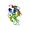
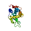
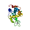
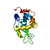
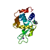
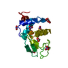
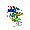
| Title | Room temperature X-ray diffraction studies of cisplatin binding to HEWL in DMSO media after 14 months of crystal storage - new refinement | ||||||
 Components Components | Lysozyme C | ||||||
 Keywords Keywords |  HYDROLASE / Re-refinement of 4G4A / Cisplatin / Metal binding HYDROLASE / Re-refinement of 4G4A / Cisplatin / Metal binding | ||||||
| Function / homology |  Function and homology information Function and homology informationLactose synthesis / Antimicrobial peptides / Neutrophil degranulation / beta-N-acetylglucosaminidase activity / cell wall macromolecule catabolic process / lysozyme / lysozyme activity / killing of cells of another organism / defense response to Gram-negative bacterium / defense response to Gram-positive bacterium ...Lactose synthesis / Antimicrobial peptides / Neutrophil degranulation / beta-N-acetylglucosaminidase activity / cell wall macromolecule catabolic process / lysozyme / lysozyme activity / killing of cells of another organism / defense response to Gram-negative bacterium / defense response to Gram-positive bacterium / defense response to bacterium / endoplasmic reticulum / extracellular space / identical protein binding / cytoplasmSimilarity search - Function | ||||||
| Biological species |  Gallus gallus (chicken) Gallus gallus (chicken) | ||||||
| Method | X-RAY DIFFRACTION / FOURIER SYNTHESIS / Resolution: 2 Å | ||||||
 Authors Authors | Shabalin, I.G. / Dauter, Z. / Jaskolski, M. / Minor, W. / Wlodawer, A. | ||||||
| Funding support |  United States, 1items United States, 1items
| ||||||
 Citation Citation | Journal: Acta Crystallogr.,Sect.D / Year: 2015 Title: Crystallography and chemistry should always go together: a cautionary tale of protein complexes with cisplatin and carboplatin. Authors: Shabalin, I. / Dauter, Z. / Jaskolski, M. / Minor, W. / Wlodawer, A. #1: Journal: Acta Crystallogr.,Sect.F / Year: 2012Title: Room-temperature X-ray diffraction studies of cisplatin and carboplatin binding to His15 of HEWL after prolonged chemical exposure. Authors: Tanley, S.W. / Schreurs, A.M. / Kroon-Batenburg, L.M. / Helliwell, J.R. | ||||||
| History |
|
- Structure visualization
| Structure viewer | Molecule: MolmilJmol/JSmol |
|---|
- Downloads & links
-Download
| PDBx/mmCIF format | 4yen.cif.gz | 69.3 KB | Display | PDBx/mmCIF format |
|---|---|---|---|---|
| PDB format | pdb4yen.ent.gz | 49.1 KB | Display | PDB format |
| PDBx/mmJSON format | 4yen.json.gz | Tree view | PDBx/mmJSON format | |
| Others |  Other downloads Other downloads |
-Validation report
| Arichive directory | https://data.pdbj.org/pub/pdb/validation_reports/ye/4yenftp://data.pdbj.org/pub/pdb/validation_reports/ye/4yen | HTTPS FTP |
|---|
-Related structure data
| Related structure data | 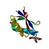 4ydxC  4yeaC  4yemC 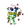 4yeoC  4g4a S: Starting model for refinement C: citing same article ( |
|---|---|
| Similar structure data |


-Links
 PDBj
PDBj


- Assembly
| Deposited unit | 
| |||||||||
|---|---|---|---|---|---|---|---|---|---|---|
| 1 |
| |||||||||
| Unit cell |
| |||||||||
| Components on special symmetry positions |
|
-Components
| #1: Protein | Mass: 14331.160 Da / Num. of mol.: 1 / Source method: isolated from a natural source / Source: (natural) Gallus gallus (chicken) / References: UniProt: P00698, lysozyme | ||||||
|---|---|---|---|---|---|---|---|
| #2: Chemical |   Mass: 195.078 Da / Num. of mol.: 2 / Source method: obtained synthetically / Formula: Pt Mass: 195.078 Da / Num. of mol.: 2 / Source method: obtained synthetically / Formula: Pt#3: Chemical | ChemComp-CL / Chloride  Mass: 35.453 Da / Num. of mol.: 5 / Source method: obtained synthetically / Formula: Cl Mass: 35.453 Da / Num. of mol.: 5 / Source method: obtained synthetically / Formula: Cl#4: Chemical | ChemComp-DMS / | Dimethyl sulfoxide  Mass: 78.133 Da / Num. of mol.: 1 / Source method: obtained synthetically / Formula: C2H6OS / Comment: DMSO, precipitant*YM Mass: 78.133 Da / Num. of mol.: 1 / Source method: obtained synthetically / Formula: C2H6OS / Comment: DMSO, precipitant*YM#5: Water | ChemComp-HOH / | Water Mass: 18.015 Da / Num. of mol.: 74 / Source method: isolated from a natural source / Formula: H2O Mass: 18.015 Da / Num. of mol.: 74 / Source method: isolated from a natural source / Formula: H2O |
-Experimental details
-Experiment
| Experiment | Method: X-RAY DIFFRACTION / Number of used crystals: 1 |
|---|
- Sample preparation
| Crystal | Density Matthews: 2.07 Å3/Da / Density % sol: 40.68 % |
|---|---|
| Crystal grow | Temperature: 295 K / Method: batch mode / pH: 4.7 Details: HEWL co-crystallized with cisplatin with 5% DMSO media in 1 mL 10% sodium chloride + 1 ml 0.04 M sodium acetate, pH 4.7 |
-Data collection
| Diffraction | Mean temperature: 295 K | ||||||||||||||||||||||||||||||||||||||||||||||||||||||||||||||||||||||||||||||||||||||||||||||||||||||||||||||||||||||||||||||||||||||||||||||||||||||||||||||||||||||||||||||||||||||||||||||||||||||||||||||||||
|---|---|---|---|---|---|---|---|---|---|---|---|---|---|---|---|---|---|---|---|---|---|---|---|---|---|---|---|---|---|---|---|---|---|---|---|---|---|---|---|---|---|---|---|---|---|---|---|---|---|---|---|---|---|---|---|---|---|---|---|---|---|---|---|---|---|---|---|---|---|---|---|---|---|---|---|---|---|---|---|---|---|---|---|---|---|---|---|---|---|---|---|---|---|---|---|---|---|---|---|---|---|---|---|---|---|---|---|---|---|---|---|---|---|---|---|---|---|---|---|---|---|---|---|---|---|---|---|---|---|---|---|---|---|---|---|---|---|---|---|---|---|---|---|---|---|---|---|---|---|---|---|---|---|---|---|---|---|---|---|---|---|---|---|---|---|---|---|---|---|---|---|---|---|---|---|---|---|---|---|---|---|---|---|---|---|---|---|---|---|---|---|---|---|---|---|---|---|---|---|---|---|---|---|---|---|---|---|---|---|---|---|
| Diffraction source | Source: ROTATING ANODE / Type: BRUKER AXS MICROSTAR / Wavelength: 1.5418 Å | ||||||||||||||||||||||||||||||||||||||||||||||||||||||||||||||||||||||||||||||||||||||||||||||||||||||||||||||||||||||||||||||||||||||||||||||||||||||||||||||||||||||||||||||||||||||||||||||||||||||||||||||||||
| Detector | Type: APEX II CCD / Detector: CCD / Date: Jan 25, 2012 | ||||||||||||||||||||||||||||||||||||||||||||||||||||||||||||||||||||||||||||||||||||||||||||||||||||||||||||||||||||||||||||||||||||||||||||||||||||||||||||||||||||||||||||||||||||||||||||||||||||||||||||||||||
| Radiation | Monochromator: confocal mirror optics / Protocol: SINGLE WAVELENGTH / Monochromatic (M) / Laue (L): M / Scattering type: x-ray | ||||||||||||||||||||||||||||||||||||||||||||||||||||||||||||||||||||||||||||||||||||||||||||||||||||||||||||||||||||||||||||||||||||||||||||||||||||||||||||||||||||||||||||||||||||||||||||||||||||||||||||||||||
| Radiation wavelength | Wavelength: 1.5418 Å / Relative weight: 1 | ||||||||||||||||||||||||||||||||||||||||||||||||||||||||||||||||||||||||||||||||||||||||||||||||||||||||||||||||||||||||||||||||||||||||||||||||||||||||||||||||||||||||||||||||||||||||||||||||||||||||||||||||||
| Reflection | Resolution: 2→50 Å / Num. all: 8602 / Num. obs: 8602 / % possible obs: 100 % / Observed criterion σ(I): -3 / Redundancy: 7.9 % / Biso Wilson estimate: 25.7 Å2 / Rmerge(I) obs: 0.086 / Rpim(I) all: 0.032 / Rrim(I) all: 0.092 / Rsym value: 0.086 / Χ2: 0.905 / Net I/av σ(I): 19.439 / Net I/σ(I): 8.8 / Num. measured all: 68208 | ||||||||||||||||||||||||||||||||||||||||||||||||||||||||||||||||||||||||||||||||||||||||||||||||||||||||||||||||||||||||||||||||||||||||||||||||||||||||||||||||||||||||||||||||||||||||||||||||||||||||||||||||||
| Reflection shell | Diffraction-ID: 1 / Rejects: 0
|
- Processing
| Software |
| |||||||||||||||||||||||||||||||||||||||||||||||||||||||||||||||||||||||||||||||||||||||||||||||||||||||||||||||||||||||||||||
|---|---|---|---|---|---|---|---|---|---|---|---|---|---|---|---|---|---|---|---|---|---|---|---|---|---|---|---|---|---|---|---|---|---|---|---|---|---|---|---|---|---|---|---|---|---|---|---|---|---|---|---|---|---|---|---|---|---|---|---|---|---|---|---|---|---|---|---|---|---|---|---|---|---|---|---|---|---|---|---|---|---|---|---|---|---|---|---|---|---|---|---|---|---|---|---|---|---|---|---|---|---|---|---|---|---|---|---|---|---|---|---|---|---|---|---|---|---|---|---|---|---|---|---|---|---|---|
| Refinement | Method to determine structure: FOURIER SYNTHESIS Starting model: 4G4A 4g4a Resolution: 2→50 Å / Cor.coef. Fo:Fc: 0.973 / Cor.coef. Fo:Fc free: 0.96 / WRfactor Rfree: 0.1507 / WRfactor Rwork: 0.1277 / FOM work R set: 0.884 / SU B: 6.737 / SU ML: 0.093 / SU R Cruickshank DPI: 0.161 / SU Rfree: 0.1334 / Cross valid method: THROUGHOUT / σ(F): 0 / ESU R: 0.161 / ESU R Free: 0.133 / Stereochemistry target values: MAXIMUM LIKELIHOOD Details: U VALUES : WITH TLS ADDED HYDROGENS HAVE BEEN ADDED IN THE RIDING POSITIONS This deposit resulted from an analysis of a number of PDB entries that contain cisplatin or carboplatin in complex ...Details: U VALUES : WITH TLS ADDED HYDROGENS HAVE BEEN ADDED IN THE RIDING POSITIONS This deposit resulted from an analysis of a number of PDB entries that contain cisplatin or carboplatin in complex with proteins, conducted in the spirit of the Terwilliger-Bricogne motto advocating continuous improvement of the macromolecular models in the PDB (Acta Cryst. D70, 2533, 2014). Since the original data frames for this structure are available at the http://rawdata.chem.uu.nl server, they have been re-processed, and the new structure factors have been used for the present re-refinement. The coordinates originally deposited as 4G4A were used as the starting point for this independent re-refinement. The new model is based on data extending to higher resolution, includes some reinterpretation of the ligands, has lower R factors, and improved statistics describing the agreement with the experimental data.
| |||||||||||||||||||||||||||||||||||||||||||||||||||||||||||||||||||||||||||||||||||||||||||||||||||||||||||||||||||||||||||||
| Solvent computation | Ion probe radii: 0.8 Å / Shrinkage radii: 0.8 Å / VDW probe radii: 1.2 Å / Solvent model: MASK | |||||||||||||||||||||||||||||||||||||||||||||||||||||||||||||||||||||||||||||||||||||||||||||||||||||||||||||||||||||||||||||
| Displacement parameters | Biso max: 69.67 Å2 / Biso mean: 28.046 Å2 / Biso min: 10.87 Å2
| |||||||||||||||||||||||||||||||||||||||||||||||||||||||||||||||||||||||||||||||||||||||||||||||||||||||||||||||||||||||||||||
| Refinement step | Cycle: final / Resolution: 2→50 Å
| |||||||||||||||||||||||||||||||||||||||||||||||||||||||||||||||||||||||||||||||||||||||||||||||||||||||||||||||||||||||||||||
| Refine LS restraints |
| |||||||||||||||||||||||||||||||||||||||||||||||||||||||||||||||||||||||||||||||||||||||||||||||||||||||||||||||||||||||||||||
| LS refinement shell | Resolution: 2→2.052 Å / Total num. of bins used: 20
| |||||||||||||||||||||||||||||||||||||||||||||||||||||||||||||||||||||||||||||||||||||||||||||||||||||||||||||||||||||||||||||
| Refinement TLS params. | Method: refined / Refine-ID: X-RAY DIFFRACTION
| |||||||||||||||||||||||||||||||||||||||||||||||||||||||||||||||||||||||||||||||||||||||||||||||||||||||||||||||||||||||||||||
| Refinement TLS group |
|
