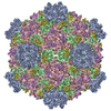
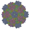
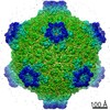
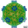
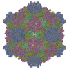
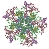
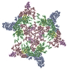
Journal: Proc Natl Acad Sci U S A / Year: 2011 Title: Atomic model of a cypovirus built from cryo-EM structure provides insight into the mechanism of mRNA capping. Authors: Lingpeng Cheng / Jingchen Sun / Kai Zhang / Zongjun Mou / Xiaoxing Huang / Gang Ji / Fei Sun / Jingqiang Zhang / Ping Zhu / Abstract: The cytoplasmic polyhedrosis virus (CPV) from the family Reoviridae belongs to a subgroup of "turreted" reoviruses, in which the mRNA capping activity occurs in a pentameric turret. We report a full ...The cytoplasmic polyhedrosis virus (CPV) from the family Reoviridae belongs to a subgroup of "turreted" reoviruses, in which the mRNA capping activity occurs in a pentameric turret. We report a full atomic model of CPV built from a 3D density map obtained using cryoelectron microscopy. The image data for the 3D reconstruction were acquired exclusively from a CCD camera. Our structure shows that the enzymatic domains of the pentameric turret of CPV are topologically conserved and that there are five unique channels connecting the guanylyltransferase and methyltransferase regions. This structural organization reveals how the channels guide nascent mRNA sequentially to guanylyltransferase, 7-N-methyltransferase, and 2'-O-methyltransferase in the turret, undergoing the highly coordinated mRNA capping activity. Furthermore, by fitting the deduced amino acid sequence of the protein VP5 to 120 large protrusion proteins on the CPV capsid shell, we confirmed that this protrusion protein is encoded by CPV RNA segment 7.
In the structure databanks used in Yorodumi, some data are registered as the other names, "COVID-19 virus" and "2019-nCoV". Here are the details of the virus and the list of structure data.
Jan 31, 2019. EMDB accession codes are about to change! (news from PDBe EMDB page)
EMDB accession codes are about to change! (news from PDBe EMDB page)
The allocation of 4 digits for EMDB accession codes will soon come to an end. Whilst these codes will remain in use, new EMDB accession codes will include an additional digit and will expand incrementally as the available range of codes is exhausted. The current 4-digit format prefixed with “EMD-” (i.e. EMD-XXXX) will advance to a 5-digit format (i.e. EMD-XXXXX), and so on. It is currently estimated that the 4-digit codes will be depleted around Spring 2019, at which point the 5-digit format will come into force.
The EM Navigator/Yorodumi systems omit the EMD- prefix.
Related info.:Q: What is EMD? / ID/Accession-code notation in Yorodumi/EM Navigator
Yorodumi is a browser for structure data from EMDB, PDB, SASBDB, etc.
This page is also the successor to EM Navigator detail page, and also detail information page/front-end page for Omokage search.
The word "yorodu" (or yorozu) is an old Japanese word meaning "ten thousand". "mi" (miru) is to see.
Related info.:EMDB / PDB / SASBDB / Comparison of 3 databanks / Yorodumi Search / Aug 31, 2016. New EM Navigator & Yorodumi / Yorodumi Papers / Jmol/JSmol / Function and homology information / Changes in new EM Navigator and Yorodumi
 Movie
Movie Controller


 Open data
 Basic information
 Components
 Keywords
 Function and homology information
 Bombyx mori cypovirus 1
 Authors
 Citation
 Structure visualization
 Downloads & links
 Other downloads


 PDBj
PDBj
 Assembly
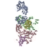

 Sample preparation
 Electron microscopy imaging
 FIELD EMISSION GUN / Accelerating voltage: 300 kV / Illumination mode: OTHER
 Processing