Resolution: 3.6→20 Å / σ(F): 0 / σ(I): 0 / Stereochemistry target values: Engh & Huber Details: positional refinement, torsion angle dynamics, individual B-factor refinement. strict 5-fold ncs restraints were maintained. NOTE: Portions of the sigma2 positioned on the ...Details: positional refinement, torsion angle dynamics, individual B-factor refinement. strict 5-fold ncs restraints were maintained. NOTE: Portions of the sigma2 positioned on the icosahedral/crystallographic 2-fold axis (for which we do not provide coordinates) were included in the refinement (occupancy 1/2 for each of two possible orientations) but were not refined. No portions at the interface with lambda1 were included.
Rfactor
Num. reflection
% reflection
Selection details
Rfree
0.2081
1428
-
random
Rwork
0.2059
-
-
-
all
0.2081
833412
-
-
obs
0.2081
831984
88.5 %
-
Refinement step
Cycle: LAST / Resolution: 3.6→20 Å
Protein
Nucleic acid
Ligand
Solvent
Total
Num. atoms
34486
0
1
0
34487
+
About Yorodumi
-
News
-
Feb 9, 2022. New format data for meta-information of EMDB entries
New format data for meta-information of EMDB entries
Version 3 of the EMDB header file is now the official format.
The previous official version 1.9 will be removed from the archive.
In the structure databanks used in Yorodumi, some data are registered as the other names, "COVID-19 virus" and "2019-nCoV". Here are the details of the virus and the list of structure data.
Jan 31, 2019. EMDB accession codes are about to change! (news from PDBe EMDB page)
EMDB accession codes are about to change! (news from PDBe EMDB page)
The allocation of 4 digits for EMDB accession codes will soon come to an end. Whilst these codes will remain in use, new EMDB accession codes will include an additional digit and will expand incrementally as the available range of codes is exhausted. The current 4-digit format prefixed with “EMD-” (i.e. EMD-XXXX) will advance to a 5-digit format (i.e. EMD-XXXXX), and so on. It is currently estimated that the 4-digit codes will be depleted around Spring 2019, at which point the 5-digit format will come into force.
The EM Navigator/Yorodumi systems omit the EMD- prefix.
Related info.:Q: What is EMD? / ID/Accession-code notation in Yorodumi/EM Navigator
Yorodumi is a browser for structure data from EMDB, PDB, SASBDB, etc.
This page is also the successor to EM Navigator detail page, and also detail information page/front-end page for Omokage search.
The word "yorodu" (or yorozu) is an old Japanese word meaning "ten thousand". "mi" (miru) is to see.
Related info.:EMDB / PDB / SASBDB / Comparison of 3 databanks / Yorodumi Search / Aug 31, 2016. New EM Navigator & Yorodumi / Yorodumi Papers / Jmol/JSmol / Function and homology information / Changes in new EM Navigator and Yorodumi
 Movie
Movie Controller


 Open data
 Basic information
 Components
 Keywords
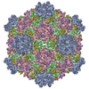
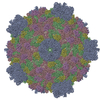
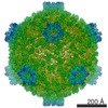
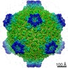
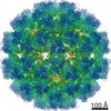
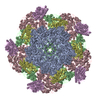
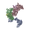
 VIRUS /
  Function and homology information
 Authors
 Citation
 Structure visualization
 Downloads & links
 Other downloads
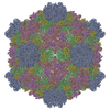
 PDBj
PDBj


 Assembly


 Mass: 65.409 Da / Num. of mol.: 1 / Source method: obtained synthetically / Formula: Zn
 Sample preparation
 / Beamline: F1 / Wavelength: 0.98
  Processing