 Movie
Movie Controller


+ Open data

- Basic information
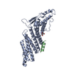
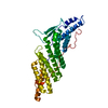
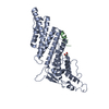
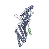
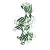
| Entry | Database: PDB / ID: 3c3q | ||||||
|---|---|---|---|---|---|---|---|
| Title | ALIX Bro1-domain:CHMIP4B co-crystal structure | ||||||
 Components Components |
| ||||||
 Keywords Keywords |  TRANSPORT PROTEIN / ALIX CHMP4B BRO1 amphipathic-helix / Cataract / Disease mutation / Protein transport / Transport / Apoptosis / Host-virus interaction TRANSPORT PROTEIN / ALIX CHMP4B BRO1 amphipathic-helix / Cataract / Disease mutation / Protein transport / Transport / Apoptosis / Host-virus interaction | ||||||
| Function / homology |  Function and homology information Function and homology informationproteinase activated receptor binding / actomyosin contractile ring assembly / ubiquitin-independent protein catabolic process via the multivesicular body sorting pathway / maintenance of lens transparency / regulation of extracellular exosome assembly / amphisome membrane / multivesicular body-lysosome fusion / viral budding / vesicle fusion with vacuole / extracellular exosome biogenesis ...proteinase activated receptor binding / actomyosin contractile ring assembly / ubiquitin-independent protein catabolic process via the multivesicular body sorting pathway / maintenance of lens transparency / regulation of extracellular exosome assembly / amphisome membrane / multivesicular body-lysosome fusion / viral budding / vesicle fusion with vacuole / extracellular exosome biogenesis / maintenance of epithelial cell apical/basal polarity / positive regulation of extracellular exosome assembly / late endosome to lysosome transport / ESCRT III complex / kinetochore microtubule / regulation of membrane permeability / late endosome to vacuole transport via multivesicular body sorting pathway / regulation of centrosome duplication / nuclear membrane reassembly / Sealing of the nuclear envelope (NE) by ESCRT-III / midbody abscission / post-translational protein targeting to endoplasmic reticulum membrane / multivesicular body sorting pathway / vesicle budding from membrane / bicellular tight junction assembly / actomyosin / membrane fission / plasma membrane repair / membrane coat / multivesicular body membrane / positive regulation of exosomal secretion / ubiquitin-dependent protein catabolic process via the multivesicular body sorting pathway / exit from mitosis / multivesicular body assembly / regulation of mitotic spindle assembly / nervous system process / Translation of Replicase and Assembly of the Replication Transcription Complex / Flemming body / nucleus organization / RIPK1-mediated regulated necrosis / Macroautophagy / mitotic metaphase chromosome alignment / viral budding via host ESCRT complex / autophagosome maturation / autophagosome membrane / mitotic cytokinesis / Uptake and function of anthrax toxins / immunological synapse / protein polymerization / bicellular tight junction / Pyroptosis / endoplasmic reticulum exit site / nuclear pore / Endosomal Sorting Complex Required For Transport (ESCRT) / multivesicular body / viral budding from plasma membrane / HCMV Late Events / regulation of autophagy / macroautophagy / Late endosomal microautophagy / Budding and maturation of HIV virion / protein homooligomerization / cytoplasmic side of plasma membrane / Regulation of necroptotic cell death / kinetochore / autophagy / calcium-dependent protein binding / extracellular vesicle / melanosome / protein transport / nuclear envelope / Translation of Replicase and Assembly of the Replication Transcription Complex / midbody / vesicle / endosome / cadherin binding / lysosomal membrane / focal adhesion / centrosome / apoptotic process / protein homodimerization activity / extracellular exosome / membrane / identical protein binding / nucleus / plasma membrane / cytosol / cytoplasmSimilarity search - Function | ||||||
| Biological species |  Homo sapiens (human) Homo sapiens (human) | ||||||
| Method | X-RAY DIFFRACTION / FOURIER SYNTHESIS / Resolution: 2.1 Å | ||||||
 Authors Authors | McCullough, J.B. / Fisher, R.D. / Whitby, F.G. / Sundquist, W.I. / Hill, C.P. | ||||||
 Citation Citation | Journal: Proc.Natl.Acad.Sci.Usa / Year: 2008 Title: ALIX-CHMP4 interactions in the human ESCRT pathway. Authors: McCullough, J. / Fisher, R.D. / Whitby, F.G. / Sundquist, W.I. / Hill, C.P. | ||||||
| History |
|
- Structure visualization
| Structure viewer | Molecule: MolmilJmol/JSmol |
|---|
- Downloads & links
-Download
| PDBx/mmCIF format | 3c3q.cif.gz | 87.5 KB | Display | PDBx/mmCIF format |
|---|---|---|---|---|
| PDB format | pdb3c3q.ent.gz | 65.5 KB | Display | PDB format |
| PDBx/mmJSON format | 3c3q.json.gz | Tree view | PDBx/mmJSON format | |
| Others |  Other downloads Other downloads |
-Validation report
| Arichive directory | https://data.pdbj.org/pub/pdb/validation_reports/c3/3c3qftp://data.pdbj.org/pub/pdb/validation_reports/c3/3c3q | HTTPS FTP |
|---|
-Related structure data


-Links
 PDBj
PDBj


- Assembly
| Deposited unit | 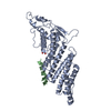
| ||||||||
|---|---|---|---|---|---|---|---|---|---|
| 1 |
| ||||||||
| Unit cell |
|
-Components
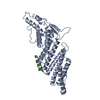
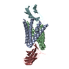
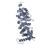
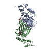
| #1: Protein | Mass: 42735.789 Da / Num. of mol.: 1 / Fragment: BRO1 domain (UNP residues 1-359) Source method: isolated from a genetically manipulated source Source: (gene. exp.) Homo sapiens (human) / Gene: PDCD6IP, AIP1, ALIX, KIAA1375 / Production host:  Escherichia coli (E. coli) / References: UniProt: Q8WUM4 Escherichia coli (E. coli) / References: UniProt: Q8WUM4 |
|---|---|
| #2: Protein/peptide | Mass: 2158.277 Da / Num. of mol.: 1 / Source method: obtained synthetically Details: CHMP4B peptide chemically synthesized. This sequence is found naturally in Homo sapiens (humans). References: UniProt: Q9H444 |
| #3: Chemical | ChemComp-GOL / Glycerol  Mass: 92.094 Da / Num. of mol.: 1 / Source method: obtained synthetically / Formula: C3H8O3 Mass: 92.094 Da / Num. of mol.: 1 / Source method: obtained synthetically / Formula: C3H8O3 |
| #4: Water | ChemComp-HOH / Water Mass: 18.015 Da / Num. of mol.: 90 / Source method: isolated from a natural source / Formula: H2O Mass: 18.015 Da / Num. of mol.: 90 / Source method: isolated from a natural source / Formula: H2O |
-Experimental details
-Experiment
| Experiment | Method: X-RAY DIFFRACTION / Number of used crystals: 1 |
|---|
- Sample preparation
| Crystal | Density Matthews: 2.72 Å3/Da / Density % sol: 54.83 % |
|---|---|
| Crystal grow | Temperature: 277 K / Method: vapor diffusion, sitting drop / pH: 6.5 Details: 15% PEG 8,000, 100mM Na MES pH 6.5, 200mM Na Acetate, VAPOR DIFFUSION, SITTING DROP, temperature 277K |
-Data collection
| Diffraction | Mean temperature: 100 K |
|---|---|
| Diffraction source | Source: ROTATING ANODE / Type: RIGAKU MICROMAX-007 HF / Wavelength: 1.5418 Å |
| Detector | Type: RIGAKU RAXIS IV / Detector: IMAGE PLATE / Date: Jan 14, 2008 / Details: VARIMAX-HR Confocal Optic |
| Radiation | Monochromator: Copper anode, varimax confocal optic / Protocol: SINGLE WAVELENGTH / Monochromatic (M) / Laue (L): M / Scattering type: x-ray |
| Radiation wavelength | Wavelength: 1.5418 Å / Relative weight: 1 |
| Reflection | Resolution: 2.1→50 Å / Num. obs: 26458 / % possible obs: 92.3 % / Observed criterion σ(F): 0 / Observed criterion σ(I): 0 |
| Reflection shell | Resolution: 2.1→2.18 Å / % possible all: 67.6 |
- Processing
| Software |
| ||||||||||||||||||||||||||||||||||||||||||||||||||||||||||||||||||||||||||||||||||||||||||||||||||||||||||||||||||||||||||||||||||||||||||||||||||||||||||||||||||||||||||
|---|---|---|---|---|---|---|---|---|---|---|---|---|---|---|---|---|---|---|---|---|---|---|---|---|---|---|---|---|---|---|---|---|---|---|---|---|---|---|---|---|---|---|---|---|---|---|---|---|---|---|---|---|---|---|---|---|---|---|---|---|---|---|---|---|---|---|---|---|---|---|---|---|---|---|---|---|---|---|---|---|---|---|---|---|---|---|---|---|---|---|---|---|---|---|---|---|---|---|---|---|---|---|---|---|---|---|---|---|---|---|---|---|---|---|---|---|---|---|---|---|---|---|---|---|---|---|---|---|---|---|---|---|---|---|---|---|---|---|---|---|---|---|---|---|---|---|---|---|---|---|---|---|---|---|---|---|---|---|---|---|---|---|---|---|---|---|---|---|---|---|---|
| Refinement | Method to determine structure: FOURIER SYNTHESIS / Resolution: 2.1→36.64 Å / Cor.coef. Fo:Fc: 0.953 / Cor.coef. Fo:Fc free: 0.922 / SU B: 16.881 / SU ML: 0.208 / Cross valid method: THROUGHOUT / σ(F): 0 / ESU R: 0.252 / ESU R Free: 0.227 / Stereochemistry target values: MAXIMUM LIKELIHOOD / Details: HYDROGENS HAVE BEEN ADDED IN THE RIDING POSITIONS
| ||||||||||||||||||||||||||||||||||||||||||||||||||||||||||||||||||||||||||||||||||||||||||||||||||||||||||||||||||||||||||||||||||||||||||||||||||||||||||||||||||||||||||
| Solvent computation | Ion probe radii: 0.8 Å / Shrinkage radii: 0.8 Å / VDW probe radii: 1.2 Å / Solvent model: MASK | ||||||||||||||||||||||||||||||||||||||||||||||||||||||||||||||||||||||||||||||||||||||||||||||||||||||||||||||||||||||||||||||||||||||||||||||||||||||||||||||||||||||||||
| Displacement parameters | Biso mean: 35.926 Å2
| ||||||||||||||||||||||||||||||||||||||||||||||||||||||||||||||||||||||||||||||||||||||||||||||||||||||||||||||||||||||||||||||||||||||||||||||||||||||||||||||||||||||||||
| Refinement step | Cycle: LAST / Resolution: 2.1→36.64 Å
| ||||||||||||||||||||||||||||||||||||||||||||||||||||||||||||||||||||||||||||||||||||||||||||||||||||||||||||||||||||||||||||||||||||||||||||||||||||||||||||||||||||||||||
| Refine LS restraints |
| ||||||||||||||||||||||||||||||||||||||||||||||||||||||||||||||||||||||||||||||||||||||||||||||||||||||||||||||||||||||||||||||||||||||||||||||||||||||||||||||||||||||||||
| LS refinement shell | Resolution: 2.1→2.154 Å / Total num. of bins used: 20
|
