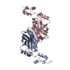
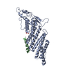
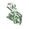
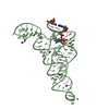
[English] 日本語
Yorodumi
- PDB-5h9m: Crystal structure of siah2 SBD domain -

+
Open data


ID or keywords:

Loading...

-
Basic information

Entry
Database: PDB / ID: 5h9m
TitleCrystal structure of siah2 SBD domain
ComponentsE3 ubiquitin-protein ligase SIAH2
KeywordsLIGASE / SBD / siah2 / Structural Genomics / Structural Genomics Consortium / SGC
Function / homology
Function and homology information


negative regulation of netrin-activated signaling pathway / protein catabolic process => GO:0030163 / Netrin-1 signaling / multicellular organism development / ubiquitin conjugating enzyme binding / small GTPase-mediated signal transduction / protein deubiquitination / regulation of protein ubiquitination / negative regulation of extrinsic apoptotic signaling pathway / RING-type E3 ubiquitin transferase ...negative regulation of netrin-activated signaling pathway / protein catabolic process => GO:0030163 / Netrin-1 signaling / multicellular organism development / ubiquitin conjugating enzyme binding / small GTPase-mediated signal transduction / protein deubiquitination / regulation of protein ubiquitination / negative regulation of extrinsic apoptotic signaling pathway / RING-type E3 ubiquitin transferase / negative regulation of cysteine-type endopeptidase activity involved in apoptotic process / negative regulation of canonical Wnt signaling pathway / regulation of circadian rhythm / protein polyubiquitination / ubiquitin-protein transferase activity / transcription corepressor activity / rhythmic process / ubiquitin protein ligase activity / Antigen processing: Ubiquitination & Proteasome degradation / ubiquitin-dependent protein catabolic process / proteasome-mediated ubiquitin-dependent protein catabolic process / amyloid fibril formation / early endosome / Ub-specific processing proteases / neuron projection / cell cycle / Amyloid fiber formation / intracellular membrane-bounded organelle / neuronal cell body / apoptotic process / negative regulation of apoptotic process / zinc ion binding / nucleoplasm / cytosol / cytoplasm
Similarity search - Function
E3 ubiquitin-protein ligase SINA-like, animal / Seven-in-absentia protein, TRAF-like domain / Sina, TRAF-like domain / Zinc finger, SIAH-type / Zinc finger SIAH-type profile. / Apoptosis, Tumor Necrosis Factor Receptor Associated Protein 2; Chain A / Apoptosis, Tumor Necrosis Factor Receptor Associated Protein 2; Chain A / TRAF-like / Zinc/RING finger domain, C3HC4 (zinc finger) / Herpes Virus-1 ...E3 ubiquitin-protein ligase SINA-like, animal / Seven-in-absentia protein, TRAF-like domain / Sina, TRAF-like domain / Zinc finger, SIAH-type / Zinc finger SIAH-type profile. / Apoptosis, Tumor Necrosis Factor Receptor Associated Protein 2; Chain A / Apoptosis, Tumor Necrosis Factor Receptor Associated Protein 2; Chain A / TRAF-like / Zinc/RING finger domain, C3HC4 (zinc finger) / Herpes Virus-1 / Zinc finger RING-type profile. / Zinc finger, RING-type / Zinc finger, RING/FYVE/PHD-type / Sandwich / 2-Layer Sandwich / Mainly Beta / Alpha Beta
Similarity search - Domain/homology
E3 ubiquitin-protein ligase SIAH2
Similarity search - Component
Biological speciesHomo sapiens (human)
MethodX-RAY DIFFRACTION / SYNCHROTRON / MOLECULAR REPLACEMENT / Resolution: 1.761 Å
AuthorsDong, A. / Zhang, Q. / Walker, J.R. / Bountra, C. / Arrowsmith, C.H. / Edwards, A.M. / Tong, Y. / Structural Genomics Consortium (SGC)
CitationJournal: to be published
Title: Crystal structure of siah2 SBD domain
Authors: Zhang, Q. / Dong, A. / Walker, J.R. / Bountra, C. / Arrowsmith, C.H. / Edwards, A.M. / Tong, Y. / Structural Genomics Consortium (SGC)
History
DepositionDec 28, 2015Deposition site: RCSB / Processing site: RCSB
Revision 1.0Feb 17, 2016Provider: repository / Type: Initial release
Revision 1.1Sep 27, 2023Group: Data collection / Database references ...Data collection / Database references / Derived calculations / Refinement description
Category: chem_comp_atom / chem_comp_bond ...chem_comp_atom / chem_comp_bond / database_2 / pdbx_initial_refinement_model / pdbx_struct_oper_list
Item: _database_2.pdbx_DOI / _database_2.pdbx_database_accession / _pdbx_struct_oper_list.symmetry_operation

-
Structure visualization

Structure viewerMolecule:
MolmilJmol/JSmol

Downloads & links

-
Assembly

Deposited unit
A: E3 ubiquitin-protein ligase SIAH2
B: E3 ubiquitin-protein ligase SIAH2
hetero molecules


Theoretical massNumber of molelcules
Total (without water)43,60315
Polymers42,7592
Non-polymers84513
Water5,513306
1


  • Idetical with deposited unit
  • defined by author&software
TypeNameSymmetry operationNumber
identity operation1_555x,y,z1
Buried area3430 Å2
ΔGint-35 kcal/mol
Surface area18450 Å2
MethodPISA
Unit cell
Length a, b, c (Å)53.624, 68.753, 102.884
Angle α, β, γ (deg.)90.00, 90.00, 90.00
Int Tables number19
Space group name H-MP212121

-
Components

-
Protein , 1 types, 2 molecules AB

#1: Protein E3 ubiquitin-protein ligase SIAH2 / Seven in absentia homolog 2 / hSiah2


Mass: 21379.400 Da / Num. of mol.: 2
Source method: isolated from a genetically manipulated source
Source: (gene. exp.) Homo sapiens (human) / Gene: SIAH2 / Plasmid: pET28-MKH8SUMO / Production host: Escherichia coli (E. coli) / Strain (production host): BL21-CodonPlus-RIL
References: UniProt: O43255, Ligases; Forming carbon-nitrogen bonds; Acid-amino-acid ligases (peptide synthases)

-
Non-polymers , 5 types, 319 molecules

#2: Chemical
ChemComp-ZN / ZINC ION


Mass: 65.409 Da / Num. of mol.: 4 / Source method: obtained synthetically / Formula: Zn
#3: Chemical ChemComp-CL / CHLORIDE ION / Chloride


Mass: 35.453 Da / Num. of mol.: 3 / Source method: obtained synthetically / Formula: Cl
#4: Chemical
ChemComp-UNX / UNKNOWN ATOM OR ION


Num. of mol.: 4 / Source method: obtained synthetically
#5: Chemical ChemComp-1PE / PENTAETHYLENE GLYCOL / PEG400 / Polyethylene glycol


Mass: 238.278 Da / Num. of mol.: 2 / Source method: obtained synthetically / Formula: C10H22O6 / Comment: precipitant*YM
#6: Water ChemComp-HOH / water / Water


Mass: 18.015 Da / Num. of mol.: 306 / Source method: isolated from a natural source / Formula: H2O

-
Experimental details

-
Experiment

ExperimentMethod: X-RAY DIFFRACTION / Number of used crystals: 1

-
Sample preparation

CrystalDensity Matthews: 2.22 Å3/Da / Density % sol: 44.54 %
Crystal growTemperature: 298 K / Method: vapor diffusion, sitting drop / pH: 7.5 / Details: 25% PEG8000, 0.2 M NaCl, 0.1 M Hepes pH7.5

-
Data collection

DiffractionMean temperature: 100 K
Diffraction sourceSource: SYNCHROTRON / Site: APS / Beamline: 19-ID / Wavelength: 0.97929 Å
DetectorType: ADSC QUANTUM 315 / Detector: CCD / Date: Jun 19, 2015
RadiationProtocol: SINGLE WAVELENGTH / Monochromatic (M) / Laue (L): M / Scattering type: x-ray
Radiation wavelengthWavelength: 0.97929 Å / Relative weight: 1
ReflectionResolution: 1.76→50.01 Å / Num. obs: 38381 / % possible obs: 100 % / Redundancy: 9.2 % / Rmerge(I) obs: 0.101 / Rpim(I) all: 0.034 / Rrim(I) all: 0.106 / Χ2: 1.143 / Net I/av σ(I): 25.746 / Net I/σ(I): 7.2 / Num. measured all: 352371
Reflection shell

Diffraction-ID: 1 / Rejects: 0

Resolution (Å)Redundancy (%)Rmerge(I) obsNum. unique allCC1/2Rpim(I) allRrim(I) allΧ2% possible all
1.76-1.796.50.87918940.720.3630.9540.68599.9
1.79-1.827.80.75118780.8280.2790.8020.696100
1.82-1.868.50.61619080.8940.2170.6540.737100
1.86-1.99.50.51918810.9340.1710.5470.771100
1.9-1.949.50.45118650.9460.1490.4760.786100
1.94-1.989.50.36619130.9610.1210.3860.81100
1.98-2.039.50.30218810.9720.10.3180.855100
2.03-2.099.50.26118950.9780.0860.2750.878100
2.09-2.159.50.22619100.9840.0750.2380.902100
2.15-2.229.50.18618960.990.0610.1960.916100
2.22-2.39.50.16919060.9910.0560.1780.941100
2.3-2.399.50.14618900.9930.0480.1540.957100
2.39-2.59.60.13319290.9940.0440.140.976100
2.5-2.639.60.11219130.9950.0370.1181.021100
2.63-2.799.60.09119090.9960.030.0961.115100
2.79-3.019.60.07719390.9970.0260.0811.257100
3.01-3.319.60.0719430.9980.0230.0741.698100
3.31-3.799.40.06519570.9980.0220.0692.384100
3.79-4.789.20.05219700.9990.0180.0552.14100
4.78-508.80.04821040.9990.0170.0511.97499.8

-
Processing

Software
NameVersionClassification
REFMAC5.8.0135refinement
SCALEPACKdata scaling
PDB_EXTRACT3.15data extraction
MOLREPphasing
ARPmodel building
RefinementMethod to determine structure: MOLECULAR REPLACEMENT
Starting model: 4C9Z
Resolution: 1.761→50.01 Å / Cor.coef. Fo:Fc: 0.962 / Cor.coef. Fo:Fc free: 0.934 / SU B: 2.747 / SU ML: 0.087 / Cross valid method: THROUGHOUT / ESU R: 0.12 / ESU R Free: 0.122 / Stereochemistry target values: MAXIMUM LIKELIHOOD / Details: HYDROGENS HAVE BEEN ADDED IN THE RIDING POSITIONS
RfactorNum. reflection% reflectionSelection details
Rfree0.22677 945 2.5 %RANDOM
Rwork0.17761 ---
obs0.17885 37374 99.7 %-
Solvent computationIon probe radii: 0.8 Å / Shrinkage radii: 0.8 Å / VDW probe radii: 1.2 Å / Solvent model: MASK
Displacement parametersBiso mean: 21.803 Å2
Baniso -1Baniso -2Baniso -3
1--0.5 Å20 Å20 Å2
2--1.52 Å2-0 Å2
3----1.02 Å2
Refinement stepCycle: LAST / Resolution: 1.761→50.01 Å
ProteinNucleic acidLigandSolventTotal
Num. atoms2935 0 39 306 3280
Refine LS restraints
Refine-IDTypeDev idealDev ideal targetNumber
X-RAY DIFFRACTIONr_bond_refined_d0.010.0193220
X-RAY DIFFRACTIONr_bond_other_d0.0020.022943
X-RAY DIFFRACTIONr_angle_refined_deg1.5061.9374394
X-RAY DIFFRACTIONr_angle_other_deg0.9323.0036766
X-RAY DIFFRACTIONr_dihedral_angle_1_deg6.3185417
X-RAY DIFFRACTIONr_dihedral_angle_2_deg38.34623.643140
X-RAY DIFFRACTIONr_dihedral_angle_3_deg11.98715501
X-RAY DIFFRACTIONr_dihedral_angle_4_deg17.8691516
X-RAY DIFFRACTIONr_chiral_restr0.0930.2479
X-RAY DIFFRACTIONr_gen_planes_refined0.0060.023754
X-RAY DIFFRACTIONr_gen_planes_other0.0020.02778
X-RAY DIFFRACTIONr_nbd_refined
X-RAY DIFFRACTIONr_nbd_other
X-RAY DIFFRACTIONr_nbtor_refined
X-RAY DIFFRACTIONr_nbtor_other
X-RAY DIFFRACTIONr_xyhbond_nbd_refined
X-RAY DIFFRACTIONr_xyhbond_nbd_other
X-RAY DIFFRACTIONr_metal_ion_refined
X-RAY DIFFRACTIONr_metal_ion_other
X-RAY DIFFRACTIONr_symmetry_vdw_refined
X-RAY DIFFRACTIONr_symmetry_vdw_other
X-RAY DIFFRACTIONr_symmetry_hbond_refined
X-RAY DIFFRACTIONr_symmetry_hbond_other
X-RAY DIFFRACTIONr_symmetry_metal_ion_refined
X-RAY DIFFRACTIONr_symmetry_metal_ion_other
X-RAY DIFFRACTIONr_mcbond_it1.3812.0331617
X-RAY DIFFRACTIONr_mcbond_other1.3792.0321616
X-RAY DIFFRACTIONr_mcangle_it2.2243.0412051
X-RAY DIFFRACTIONr_mcangle_other2.2243.0412052
X-RAY DIFFRACTIONr_scbond_it1.8452.2411603
X-RAY DIFFRACTIONr_scbond_other1.8442.2421604
X-RAY DIFFRACTIONr_scangle_it
X-RAY DIFFRACTIONr_scangle_other2.9793.2732343
X-RAY DIFFRACTIONr_long_range_B_refined5.12117.2813697
X-RAY DIFFRACTIONr_long_range_B_other5.0317.1263651
X-RAY DIFFRACTIONr_rigid_bond_restr
X-RAY DIFFRACTIONr_sphericity_free
X-RAY DIFFRACTIONr_sphericity_bonded
LS refinement shellResolution: 1.761→1.806 Å / Total num. of bins used: 20
RfactorNum. reflection% reflection
Rfree0.24 65 -
Rwork0.256 2633 -
obs--96.08 %

+
About Yorodumi

-
News

-
Feb 9, 2022. New format data for meta-information of EMDB entries

New format data for meta-information of EMDB entries

  • Version 3 of the EMDB header file is now the official format.
  • The previous official version 1.9 will be removed from the archive.

Related info.:EMDB header

External links:wwPDB to switch to version 3 of the EMDB data model

-
Aug 12, 2020. Covid-19 info

Covid-19 info

URL: https://pdbj.org/emnavi/covid19.php

New page: Covid-19 featured information page in EM Navigator.

Related info.:Covid-19 info / Mar 5, 2020. Novel coronavirus structure data

+
Mar 5, 2020. Novel coronavirus structure data

Novel coronavirus structure data

Related info.:Yorodumi Speices / Aug 12, 2020. Covid-19 info

External links:COVID-19 featured content - PDBj / Molecule of the Month (242):Coronavirus Proteases

+
Jan 31, 2019. EMDB accession codes are about to change! (news from PDBe EMDB page)

EMDB accession codes are about to change! (news from PDBe EMDB page)

  • The allocation of 4 digits for EMDB accession codes will soon come to an end. Whilst these codes will remain in use, new EMDB accession codes will include an additional digit and will expand incrementally as the available range of codes is exhausted. The current 4-digit format prefixed with “EMD-” (i.e. EMD-XXXX) will advance to a 5-digit format (i.e. EMD-XXXXX), and so on. It is currently estimated that the 4-digit codes will be depleted around Spring 2019, at which point the 5-digit format will come into force.
  • The EM Navigator/Yorodumi systems omit the EMD- prefix.

Related info.:Q: What is EMD? / ID/Accession-code notation in Yorodumi/EM Navigator

External links:EMDB Accession Codes are Changing Soon! / Contact to PDBj

+
Jul 12, 2017. Major update of PDB

Major update of PDB

  • wwPDB released updated PDB data conforming to the new PDBx/mmCIF dictionary.
  • This is a major update changing the version number from 4 to 5, and with Remediation, in which all the entries are updated.
  • In this update, many items about electron microscopy experimental information are reorganized (e.g. em_software).
  • Now, EM Navigator and Yorodumi are based on the updated data.

External links:wwPDB Remediation / Enriched Model Files Conforming to OneDep Data Standards Now Available in the PDB FTP Archive

-
Yorodumi

Thousand views of thousand structures

  • Yorodumi is a browser for structure data from EMDB, PDB, SASBDB, etc.
  • This page is also the successor to EM Navigator detail page, and also detail information page/front-end page for Omokage search.
  • The word "yorodu" (or yorozu) is an old Japanese word meaning "ten thousand". "mi" (miru) is to see.

Related info.:EMDB / PDB / SASBDB / Comparison of 3 databanks / Yorodumi Search / Aug 31, 2016. New EM Navigator & Yorodumi / Yorodumi Papers / Jmol/JSmol / Function and homology information / Changes in new EM Navigator and Yorodumi

Read more