 Movie
Movie Controller

[English] 日本語
 Yorodumi
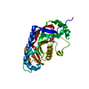
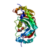
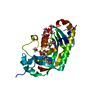
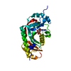
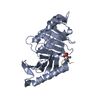
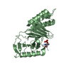
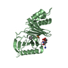
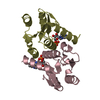
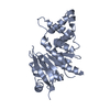
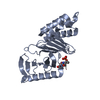
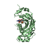
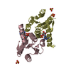
- PDB-2yoz: Catalytic domain of mouse 2',3'-cyclic nucleotide 3'- phosphodies... -
+ Open data

- Basic information
| Entry | Database: PDB / ID: 2yoz | |||||||||
|---|---|---|---|---|---|---|---|---|---|---|
| Title | Catalytic domain of mouse 2',3'-cyclic nucleotide 3'- phosphodiesterase, crystallized with 2'-AMPS | |||||||||
 Components Components | 2,3-CYCLIC NUCLEOTIDE 3'-PHOSPHODIESTERASE | |||||||||
 Keywords Keywords |  HYDROLASE / MYELIN / NERVOUS SYSTEM HYDROLASE / MYELIN / NERVOUS SYSTEM | |||||||||
| Function / homology |  Function and homology information Function and homology informationcyclic nucleotide catabolic process / 2',3'-cyclic-nucleotide 3'-phosphodiesterase / 2',3'-cyclic-nucleotide 3'-phosphodiesterase activity / myelin sheath abaxonal region / myelin sheath adaxonal region / cyclic nucleotide binding / regulation of mitochondrial membrane permeability / oligodendrocyte differentiation / pseudopodium / microvillus ...cyclic nucleotide catabolic process / 2',3'-cyclic-nucleotide 3'-phosphodiesterase / 2',3'-cyclic-nucleotide 3'-phosphodiesterase activity / myelin sheath abaxonal region / myelin sheath adaxonal region / cyclic nucleotide binding / regulation of mitochondrial membrane permeability / oligodendrocyte differentiation / pseudopodium / microvillus / forebrain development / axonogenesis / adult locomotory behavior / cell projection / response to toxic substance / melanosome / myelin sheath / mitochondrial inner membrane / microtubule / mitochondrial outer membrane / response to lipopolysaccharide / perinuclear region of cytoplasm / extracellular space / RNA binding / membrane / cytoplasmSimilarity search - Function | |||||||||
| Biological species |  MUS MUSCULUS (house mouse) MUS MUSCULUS (house mouse) | |||||||||
| Method | X-RAY DIFFRACTION / SYNCHROTRON / MOLECULAR REPLACEMENT / Resolution: 2.1 Å | |||||||||
 Authors Authors | Myllykoski, M. / Raasakka, A. / Lehtimaki, M. / Han, H. / Kursula, P. | |||||||||
 Citation Citation | Journal: J.Mol.Biol. / Year: 2013 Title: Crystallographic Analysis of the Reaction Cycle of 2',3'-Cyclic Nucleotide 3'-Phosphodiesterase, a Unique Member of the 2H Phosphoesterase Family Authors: Myllykoski, M. / Raasakka, A. / Lehtimaki, M. / Han, H. / Kursula, I. / Kursula, P. | |||||||||
| History |
|
- Structure visualization
| Structure viewer | Molecule: MolmilJmol/JSmol |
|---|
- Downloads & links
-Download
| PDBx/mmCIF format | 2yoz.cif.gz | 138.8 KB | Display | PDBx/mmCIF format |
|---|---|---|---|---|
| PDB format | pdb2yoz.ent.gz | 110.2 KB | Display | PDB format |
| PDBx/mmJSON format | 2yoz.json.gz | Tree view | PDBx/mmJSON format | |
| Others |  Other downloads Other downloads |
-Validation report
| Arichive directory | https://data.pdbj.org/pub/pdb/validation_reports/yo/2yozftp://data.pdbj.org/pub/pdb/validation_reports/yo/2yoz | HTTPS FTP |
|---|
-Related structure data
| Related structure data |  2yp0C  2ypcC  2ypeC 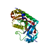 2yphC  2yq9C 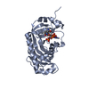 3zbrC 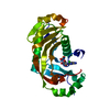 3zbsC  3zbzC 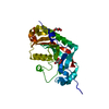 2xmiS S: Starting model for refinement C: citing same article ( |
|---|---|
| Similar structure data |


-Links
 PDBj
PDBj- Assembly
| Deposited unit | 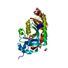
| ||||||||
|---|---|---|---|---|---|---|---|---|---|
| 1 |
| ||||||||
| Unit cell |
|
-Components
-Protein , 1 types, 1 molecules A
| #1: Protein | Mass: 24293.928 Da / Num. of mol.: 1 / Fragment: C-TERMINAL CATALYTIC DOMAIN, RESIDUES 159-378 Source method: isolated from a genetically manipulated source Source: (gene. exp.) MUS MUSCULUS (house mouse) / Plasmid: PTH 27 / Production host:  ESCHERICHIA COLI (E. coli) / Strain (production host): BL21(DE3) / Variant (production host): ROSETTA ESCHERICHIA COLI (E. coli) / Strain (production host): BL21(DE3) / Variant (production host): ROSETTAReferences: UniProt: P16330, 2',3'-cyclic-nucleotide 3'-phosphodiesterase |
|---|
-Non-polymers , 6 types, 105 molecules 


| #2: Chemical | ChemComp-OVE /  Mass: 363.287 Da / Num. of mol.: 1 / Source method: obtained synthetically / Formula: C10H14N5O6PS Mass: 363.287 Da / Num. of mol.: 1 / Source method: obtained synthetically / Formula: C10H14N5O6PS | ||||
|---|---|---|---|---|---|
| #3: Chemical | ChemComp-GOL / Glycerol Mass: 92.094 Da / Num. of mol.: 1 / Source method: obtained synthetically / Formula: C3H8O3 Mass: 92.094 Da / Num. of mol.: 1 / Source method: obtained synthetically / Formula: C3H8O3 | ||||
| #4: Chemical | ChemComp-1PE / Polyethylene glycol Mass: 238.278 Da / Num. of mol.: 1 / Source method: obtained synthetically / Formula: C10H22O6 / Comment: precipitant*YM Mass: 238.278 Da / Num. of mol.: 1 / Source method: obtained synthetically / Formula: C10H22O6 / Comment: precipitant*YM | ||||
| #5: Chemical | ChemComp-ACT / Acetate Mass: 59.044 Da / Num. of mol.: 4 / Source method: obtained synthetically / Formula: C2H3O2 Mass: 59.044 Da / Num. of mol.: 4 / Source method: obtained synthetically / Formula: C2H3O2#6: Chemical | ChemComp-CL / | Chloride Mass: 35.453 Da / Num. of mol.: 1 / Source method: obtained synthetically / Formula: Cl Mass: 35.453 Da / Num. of mol.: 1 / Source method: obtained synthetically / Formula: Cl#7: Water | ChemComp-HOH / | WaterMass: 18.015 Da / Num. of mol.: 97 / Source method: isolated from a natural source / Formula: H2O |
-Details
| Sequence details | N-TERMINAL GLYCINE REMAINS FROM TEV-CLEAVAGE OF EXPRESSION TAG. RESIDUE NUMBERING IS ACCORDING TO ...N-TERMINAL GLYCINE REMAINS FROM TEV-CLEAVAGE OF EXPRESSION |
|---|
-Experimental details
-Experiment
| Experiment | Method: X-RAY DIFFRACTION / Number of used crystals: 1 |
|---|
- Sample preparation
| Crystal | Density Matthews: 1.9 Å3/Da / Density % sol: 35.1 % / Description: NONE |
|---|---|
| Crystal grow | Temperature: 277 K / pH: 4 Details: 250 UM PROTEIN AND 10 MM 2, 3(SP)-CYCLIC-AMPS MIXED IN 0.5 PLUS 0.5 DROPS WITH 30% PEG4000 AND 50 MM ACETATE (1:1 MIX OF PH 3 AND PH 5) IN 4C TEMP |
-Data collection
| Diffraction | Mean temperature: 100 K |
|---|---|
| Diffraction source | Source: SYNCHROTRON / Site: EMBL/DESY, HAMBURG  / Beamline: X11 / Wavelength: 0.815 / Beamline: X11 / Wavelength: 0.815 |
| Detector | Type: MAR555 FLAT PANEL / Detector: IMAGE PLATE / Date: Apr 3, 2012 |
| Radiation | Protocol: SINGLE WAVELENGTH / Monochromatic (M) / Laue (L): M / Scattering type: x-ray |
| Radiation wavelength | Wavelength: 0.815 Å / Relative weight: 1 |
| Reflection | Resolution: 2.1→19.1 Å / Num. obs: 10896 / % possible obs: 99.4 % / Observed criterion σ(I): -3 / Redundancy: 3.5 % / Biso Wilson estimate: 28.21 Å2 / Rmerge(I) obs: 0.08 / Net I/σ(I): 11.7 |
| Reflection shell | Resolution: 2.1→2.15 Å / Redundancy: 2.7 % / Rmerge(I) obs: 0.52 / Mean I/σ(I) obs: 2 / % possible all: 98.6 |
- Processing
| Software |
| ||||||||||||||||||||||||||||||||||||||||||||||||||||||||||||||||||||||||||||||||||||||||||||||||||||||||||||||||||||||||||||||||||||||||||||||||||||||||||||||||||||||||||||||||||||||||||||||||||||||||
|---|---|---|---|---|---|---|---|---|---|---|---|---|---|---|---|---|---|---|---|---|---|---|---|---|---|---|---|---|---|---|---|---|---|---|---|---|---|---|---|---|---|---|---|---|---|---|---|---|---|---|---|---|---|---|---|---|---|---|---|---|---|---|---|---|---|---|---|---|---|---|---|---|---|---|---|---|---|---|---|---|---|---|---|---|---|---|---|---|---|---|---|---|---|---|---|---|---|---|---|---|---|---|---|---|---|---|---|---|---|---|---|---|---|---|---|---|---|---|---|---|---|---|---|---|---|---|---|---|---|---|---|---|---|---|---|---|---|---|---|---|---|---|---|---|---|---|---|---|---|---|---|---|---|---|---|---|---|---|---|---|---|---|---|---|---|---|---|---|---|---|---|---|---|---|---|---|---|---|---|---|---|---|---|---|---|---|---|---|---|---|---|---|---|---|---|---|---|---|---|---|---|
| Refinement | Method to determine structure: MOLECULAR REPLACEMENT Starting model: PDB ENTRY 2XMI Resolution: 2.1→16.999 Å / SU ML: 0.27 / σ(F): 2 / Phase error: 24.59 / Stereochemistry target values: ML Details: HYDROGENS WERE INCLUDED IN RIDING POSITION. RESIDUES 158-160 AND 206-212 WERE EXCLUDED DUE TO INSUFFICIENT ELECTRON DENSITY.
| ||||||||||||||||||||||||||||||||||||||||||||||||||||||||||||||||||||||||||||||||||||||||||||||||||||||||||||||||||||||||||||||||||||||||||||||||||||||||||||||||||||||||||||||||||||||||||||||||||||||||
| Solvent computation | Shrinkage radii: 0.9 Å / VDW probe radii: 1.11 Å / Solvent model: FLAT BULK SOLVENT MODEL | ||||||||||||||||||||||||||||||||||||||||||||||||||||||||||||||||||||||||||||||||||||||||||||||||||||||||||||||||||||||||||||||||||||||||||||||||||||||||||||||||||||||||||||||||||||||||||||||||||||||||
| Displacement parameters | Biso mean: 33.9 Å2 | ||||||||||||||||||||||||||||||||||||||||||||||||||||||||||||||||||||||||||||||||||||||||||||||||||||||||||||||||||||||||||||||||||||||||||||||||||||||||||||||||||||||||||||||||||||||||||||||||||||||||
| Refinement step | Cycle: LAST / Resolution: 2.1→16.999 Å
| ||||||||||||||||||||||||||||||||||||||||||||||||||||||||||||||||||||||||||||||||||||||||||||||||||||||||||||||||||||||||||||||||||||||||||||||||||||||||||||||||||||||||||||||||||||||||||||||||||||||||
| Refine LS restraints |
| ||||||||||||||||||||||||||||||||||||||||||||||||||||||||||||||||||||||||||||||||||||||||||||||||||||||||||||||||||||||||||||||||||||||||||||||||||||||||||||||||||||||||||||||||||||||||||||||||||||||||
| LS refinement shell |
| ||||||||||||||||||||||||||||||||||||||||||||||||||||||||||||||||||||||||||||||||||||||||||||||||||||||||||||||||||||||||||||||||||||||||||||||||||||||||||||||||||||||||||||||||||||||||||||||||||||||||
| Refinement TLS params. | Method: refined / Refine-ID: X-RAY DIFFRACTION
| ||||||||||||||||||||||||||||||||||||||||||||||||||||||||||||||||||||||||||||||||||||||||||||||||||||||||||||||||||||||||||||||||||||||||||||||||||||||||||||||||||||||||||||||||||||||||||||||||||||||||
| Refinement TLS group |
|
