Mitotic Telophase/Cytokinesis / regulation of protein localization to cell cortex / Mitotic Metaphase/Anaphase Transition / Golgi inheritance / synaptonemal complex disassembly / Activation of NIMA Kinases NEK9, NEK6, NEK7 / homologous chromosome segregation / polo kinase / nuclear membrane disassembly / mitotic nuclear membrane disassembly ...Mitotic Telophase/Cytokinesis / regulation of protein localization to cell cortex / Mitotic Metaphase/Anaphase Transition / Golgi inheritance / synaptonemal complex disassembly / Activation of NIMA Kinases NEK9, NEK6, NEK7 / homologous chromosome segregation / polo kinase / nuclear membrane disassembly / mitotic nuclear membrane disassembly / protein localization to nuclear envelope / Phosphorylation of Emi1 / metaphase/anaphase transition of mitotic cell cycle / synaptonemal complex / female meiosis chromosome segregation / Phosphorylation of the APC/C / regulation of protein binding / anaphase-promoting complex binding / outer kinetochore / negative regulation of cyclin-dependent protein serine/threonine kinase activity / positive regulation of ubiquitin protein ligase activity / regulation of mitotic spindle assembly / microtubule bundle formation / Polo-like kinase mediated events / Golgi Cisternae Pericentriolar Stack Reorganization / mitotic chromosome condensation / regulation of mitotic metaphase/anaphase transition / sister chromatid cohesion / positive regulation of ubiquitin-protein transferase activity / centrosome cycle / double-strand break repair via alternative nonhomologous end joining / regulation of mitotic cell cycle phase transition / mitotic spindle assembly checkpoint signaling / mitotic spindle pole / regulation of anaphase-promoting complex-dependent catabolic process / mitotic G2 DNA damage checkpoint signaling / mitotic sister chromatid segregation / establishment of mitotic spindle orientation / positive regulation of proteolysis / mitotic cytokinesis / centriolar satellite / spindle midzone / negative regulation of double-strand break repair via homologous recombination / Amplification of signal from unattached kinetochores via a MAD2 inhibitory signal / Cyclin A/B1/B2 associated events during G2/M transition / Mitotic Prometaphase / EML4 and NUDC in mitotic spindle formation / Loss of Nlp from mitotic centrosomes / Loss of proteins required for interphase microtubule organization from the centrosome / Recruitment of mitotic centrosome proteins and complexes / protein localization to chromatin / Resolution of Sister Chromatid Cohesion / Recruitment of NuMA to mitotic centrosomes / Anchoring of the basal body to the plasma membrane / regulation of mitotic cell cycle / centriole / AURKA Activation by TPX2 / Condensation of Prophase Chromosomes / mitotic spindle organization / positive regulation of peptidyl-threonine phosphorylation / regulation of cytokinesis / RHO GTPases Activate Formins / protein destabilization / APC/C:Cdh1 mediated degradation of Cdc20 and other APC/C:Cdh1 targeted proteins in late mitosis/early G1 / establishment of protein localization / kinetochore / spindle pole / spindle / positive regulation of protein localization to nucleus / Separation of Sister Chromatids / The role of GTSE1 in G2/M progression after G2 checkpoint / G2/M transition of mitotic cell cycle / microtubule cytoskeleton / Regulation of PLK1 Activity at G2/M Transition / double-strand break repair / positive regulation of proteasomal ubiquitin-dependent protein catabolic process / mitotic cell cycle / midbody / microtubule binding / peptidyl-serine phosphorylation / protein ubiquitination / regulation of cell cycle / protein kinase activity / protein phosphorylation / protein serine kinase activity / protein serine/threonine kinase activity / centrosome / chromatin / negative regulation of apoptotic process / protein kinase binding / negative regulation of transcription by RNA polymerase II / magnesium ion binding / nucleoplasm / ATP binding / identical protein binding / nucleus / cytosol / cytoplasm Similarity search - Function
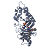
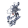
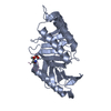
POLO box domain / Polo-like kinase 1, catalytic domain / Second polo-box domain / First polo-box domain / POLO box domain superfamily / POLO box duplicated region / POLO box domain / POLO box domain profile. / Arylsulfatase, C-terminal domain / Serine/threonine-protein kinase, active site ...POLO box domain / Polo-like kinase 1, catalytic domain / Second polo-box domain / First polo-box domain / POLO box domain superfamily / POLO box duplicated region / POLO box domain / POLO box domain profile. / Arylsulfatase, C-terminal domain / Serine/threonine-protein kinase, active site / Serine/Threonine protein kinases active-site signature. / Protein kinase domain / Serine/Threonine protein kinases, catalytic domain / Protein kinase, ATP binding site / Protein kinases ATP-binding region signature. / Protein kinase domain profile. / Protein kinase domain / Protein kinase-like domain superfamily / 2-Layer Sandwich / Alpha Beta Similarity search - Domain/homology
 Movie
Movie Controller


 Open data
 Basic information
 Components
 Keywords
 TRANSFERASE /
  Function and homology information
 Authors
 Citation
 Structure visualization
 Downloads & links
 Other downloads


 PDBj
PDBj


 Assembly
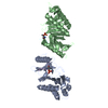

 Mass: 18.015 Da / Num. of mol.: 275 / Source method: isolated from a natural source / Formula: H2O
 Sample preparation
 / Beamline: PX14.1 / Wavelength: 1.244
  Processing