 Movie
Movie Controller

[English] 日本語
 Yorodumi
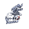
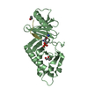
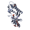
- PDB-4x9w: PLK-1 polo-box domain in complex with Bioactive Imidazolium-conta... -
+ Open data

- Basic information
| Entry | Database: PDB / ID: 4x9w | |||||||||
|---|---|---|---|---|---|---|---|---|---|---|
| Title | PLK-1 polo-box domain in complex with Bioactive Imidazolium-containing phosphopeptide macrocycle 4C | |||||||||
 Components Components |
| |||||||||
 Keywords Keywords | TRANSFERASE/TRANSFERASE INHIBITOR / TRANSFERASE-TRANSFERASE INHIBITOR complex | |||||||||
| Function / homology |  Function and homology information Function and homology informationMitotic Telophase/Cytokinesis / regulation of protein localization to cell cortex / Mitotic Metaphase/Anaphase Transition / Golgi inheritance / synaptonemal complex disassembly / Activation of NIMA Kinases NEK9, NEK6, NEK7 / homologous chromosome segregation /  polo kinase / nuclear membrane disassembly / mitotic nuclear membrane disassembly ...Mitotic Telophase/Cytokinesis / regulation of protein localization to cell cortex / Mitotic Metaphase/Anaphase Transition / Golgi inheritance / synaptonemal complex disassembly / Activation of NIMA Kinases NEK9, NEK6, NEK7 / homologous chromosome segregation / polo kinase / nuclear membrane disassembly / mitotic nuclear membrane disassembly / protein localization to nuclear envelope / Phosphorylation of Emi1 / metaphase/anaphase transition of mitotic cell cycle / synaptonemal complex / female meiosis chromosome segregation / Phosphorylation of the APC/C / regulation of protein binding / anaphase-promoting complex binding / outer kinetochore / negative regulation of cyclin-dependent protein serine/threonine kinase activity / positive regulation of ubiquitin protein ligase activity / regulation of mitotic spindle assembly / microtubule bundle formation / Polo-like kinase mediated events / Golgi Cisternae Pericentriolar Stack Reorganization / mitotic chromosome condensation / regulation of mitotic metaphase/anaphase transition / sister chromatid cohesion / positive regulation of ubiquitin-protein transferase activity / centrosome cycle / double-strand break repair via alternative nonhomologous end joining / regulation of mitotic cell cycle phase transition / mitotic spindle assembly checkpoint signaling / mitotic spindle pole / regulation of anaphase-promoting complex-dependent catabolic process / mitotic G2 DNA damage checkpoint signaling / mitotic sister chromatid segregation / establishment of mitotic spindle orientation / positive regulation of proteolysis / mitotic cytokinesis / centriolar satellite / spindle midzone / negative regulation of double-strand break repair via homologous recombination / Amplification of signal from unattached kinetochores via a MAD2 inhibitory signal / Cyclin A/B1/B2 associated events during G2/M transition / Mitotic Prometaphase / EML4 and NUDC in mitotic spindle formation / Loss of Nlp from mitotic centrosomes / Loss of proteins required for interphase microtubule organization from the centrosome / Recruitment of mitotic centrosome proteins and complexes / protein localization to chromatin / Resolution of Sister Chromatid Cohesion / Recruitment of NuMA to mitotic centrosomes / Anchoring of the basal body to the plasma membrane / regulation of mitotic cell cycle / centriole / AURKA Activation by TPX2 / Condensation of Prophase Chromosomes / mitotic spindle organization / positive regulation of peptidyl-threonine phosphorylation / regulation of cytokinesis / RHO GTPases Activate Formins / protein destabilization / APC/C:Cdh1 mediated degradation of Cdc20 and other APC/C:Cdh1 targeted proteins in late mitosis/early G1 / establishment of protein localization / kinetochore / spindle pole / spindle / positive regulation of protein localization to nucleus / Separation of Sister Chromatids / The role of GTSE1 in G2/M progression after G2 checkpoint / G2/M transition of mitotic cell cycle / microtubule cytoskeleton / Regulation of PLK1 Activity at G2/M Transition / double-strand break repair / positive regulation of proteasomal ubiquitin-dependent protein catabolic process / mitotic cell cycle / midbody / microtubule binding / peptidyl-serine phosphorylation / protein ubiquitination / regulation of cell cycle / protein kinase activity / protein phosphorylation / protein serine kinase activity / protein serine/threonine kinase activity / centrosome / chromatin / negative regulation of apoptotic process / protein kinase binding / negative regulation of transcription by RNA polymerase II / magnesium ion binding / nucleoplasm / ATP binding / identical protein binding / nucleus / cytosol / cytoplasm polo kinase / nuclear membrane disassembly / mitotic nuclear membrane disassembly ...Mitotic Telophase/Cytokinesis / regulation of protein localization to cell cortex / Mitotic Metaphase/Anaphase Transition / Golgi inheritance / synaptonemal complex disassembly / Activation of NIMA Kinases NEK9, NEK6, NEK7 / homologous chromosome segregation / polo kinase / nuclear membrane disassembly / mitotic nuclear membrane disassembly / protein localization to nuclear envelope / Phosphorylation of Emi1 / metaphase/anaphase transition of mitotic cell cycle / synaptonemal complex / female meiosis chromosome segregation / Phosphorylation of the APC/C / regulation of protein binding / anaphase-promoting complex binding / outer kinetochore / negative regulation of cyclin-dependent protein serine/threonine kinase activity / positive regulation of ubiquitin protein ligase activity / regulation of mitotic spindle assembly / microtubule bundle formation / Polo-like kinase mediated events / Golgi Cisternae Pericentriolar Stack Reorganization / mitotic chromosome condensation / regulation of mitotic metaphase/anaphase transition / sister chromatid cohesion / positive regulation of ubiquitin-protein transferase activity / centrosome cycle / double-strand break repair via alternative nonhomologous end joining / regulation of mitotic cell cycle phase transition / mitotic spindle assembly checkpoint signaling / mitotic spindle pole / regulation of anaphase-promoting complex-dependent catabolic process / mitotic G2 DNA damage checkpoint signaling / mitotic sister chromatid segregation / establishment of mitotic spindle orientation / positive regulation of proteolysis / mitotic cytokinesis / centriolar satellite / spindle midzone / negative regulation of double-strand break repair via homologous recombination / Amplification of signal from unattached kinetochores via a MAD2 inhibitory signal / Cyclin A/B1/B2 associated events during G2/M transition / Mitotic Prometaphase / EML4 and NUDC in mitotic spindle formation / Loss of Nlp from mitotic centrosomes / Loss of proteins required for interphase microtubule organization from the centrosome / Recruitment of mitotic centrosome proteins and complexes / protein localization to chromatin / Resolution of Sister Chromatid Cohesion / Recruitment of NuMA to mitotic centrosomes / Anchoring of the basal body to the plasma membrane / regulation of mitotic cell cycle / centriole / AURKA Activation by TPX2 / Condensation of Prophase Chromosomes / mitotic spindle organization / positive regulation of peptidyl-threonine phosphorylation / regulation of cytokinesis / RHO GTPases Activate Formins / protein destabilization / APC/C:Cdh1 mediated degradation of Cdc20 and other APC/C:Cdh1 targeted proteins in late mitosis/early G1 / establishment of protein localization / kinetochore / spindle pole / spindle / positive regulation of protein localization to nucleus / Separation of Sister Chromatids / The role of GTSE1 in G2/M progression after G2 checkpoint / G2/M transition of mitotic cell cycle / microtubule cytoskeleton / Regulation of PLK1 Activity at G2/M Transition / double-strand break repair / positive regulation of proteasomal ubiquitin-dependent protein catabolic process / mitotic cell cycle / midbody / microtubule binding / peptidyl-serine phosphorylation / protein ubiquitination / regulation of cell cycle / protein kinase activity / protein phosphorylation / protein serine kinase activity / protein serine/threonine kinase activity / centrosome / chromatin / negative regulation of apoptotic process / protein kinase binding / negative regulation of transcription by RNA polymerase II / magnesium ion binding / nucleoplasm / ATP binding / identical protein binding / nucleus / cytosol / cytoplasmSimilarity search - Function | |||||||||
| Biological species |  Homo sapiens (human) Homo sapiens (human)synthetic construct (others) | |||||||||
| Method | X-RAY DIFFRACTION / SYNCHROTRON / MOLECULAR REPLACEMENT / Resolution: 1.798 Å | |||||||||
 Authors Authors | Grant, R.A. / Qian, W.-J. / Yaffe, M.B. / Burke, T.R. | |||||||||
 Citation Citation | Journal: Biopolymers / Year: 2015 Title: Neighbor-directed histidine N ( tau )-alkylation: A route to imidazolium-containing phosphopeptide macrocycles. Authors: Qian, W.J. / Park, J.E. / Grant, R. / Lai, C.C. / Kelley, J.A. / Yaffe, M.B. / Lee, K.S. / Burke, T.R. | |||||||||
| History |
|
- Structure visualization
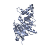
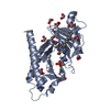
| Structure viewer | Molecule: MolmilJmol/JSmol |
|---|
- Downloads & links
-Download
| PDBx/mmCIF format | 4x9w.cif.gz | 109 KB | Display | PDBx/mmCIF format |
|---|---|---|---|---|
| PDB format | pdb4x9w.ent.gz | 84.2 KB | Display | PDB format |
| PDBx/mmJSON format | 4x9w.json.gz | Tree view | PDBx/mmJSON format | |
| Others |  Other downloads Other downloads |
-Validation report
| Arichive directory | https://data.pdbj.org/pub/pdb/validation_reports/x9/4x9wftp://data.pdbj.org/pub/pdb/validation_reports/x9/4x9w | HTTPS FTP |
|---|
-Related structure data
| Related structure data |  4x9rC  4x9vC  3rq7S C: citing same article ( S: Starting model for refinement |
|---|---|
| Similar structure data |


-Links
 PDBj
PDBj


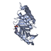
- Assembly
| Deposited unit | 
| ||||||||
|---|---|---|---|---|---|---|---|---|---|
| 1 |
| ||||||||
| Unit cell |
|
-Components
| #1: Protein | Mass: 27285.158 Da / Num. of mol.: 1 / Fragment: POLO box domain residues 371-603 Source method: isolated from a genetically manipulated source Source: (gene. exp.) Homo sapiens (human) / Gene: PLK1, PLK / Production host:  Escherichia coli (E. coli) / References: UniProt: P53350, polo kinase Escherichia coli (E. coli) / References: UniProt: P53350, polo kinase |
|---|---|
| #2: Protein/peptide | Mass: 909.081 Da / Num. of mol.: 1 / Source method: obtained synthetically / Source: (synth.) synthetic construct (others) |
| #3: Water | ChemComp-HOH / Water Mass: 18.015 Da / Num. of mol.: 73 / Source method: isolated from a natural source / Formula: H2O Mass: 18.015 Da / Num. of mol.: 73 / Source method: isolated from a natural source / Formula: H2O |
-Experimental details
-Experiment
| Experiment | Method: X-RAY DIFFRACTION / Number of used crystals: 1 |
|---|
- Sample preparation
| Crystal | Density Matthews: 1.92 Å3/Da / Density % sol: 36.02 % |
|---|---|
| Crystal grow | Temperature: 291 K / Method: vapor diffusion / pH: 8 Details: Frozen stocks of protein at 37 mg/mL in 10 mM TRIS pH8, 0.5 M NaCl, 10 mM DTT were thawed and diluted to 10 mg/ml with the same buffer. Complexes with each of three macrocycle compounds (3b, ...Details: Frozen stocks of protein at 37 mg/mL in 10 mM TRIS pH8, 0.5 M NaCl, 10 mM DTT were thawed and diluted to 10 mg/ml with the same buffer. Complexes with each of three macrocycle compounds (3b, 3c and 4b) were prepared by adding 100 mM stocks of the macrocycle in DMSO directly to the diluted protein to achieve a final concentration of 1 mM. Crystals were grown by hanging drop vapor diffusion, with drops made by mixing equal volumes of protein-macrocycle complex and well solution containing 2-6% PEG-3350. Crystals were cryo-protected by quickly dipping in a solution of 37.5% ethylene glycol in well solution and frozen in liquid nitrogen. |
-Data collection
| Diffraction | Mean temperature: 100 K | |||||||||||||||||||||||||||||||||||||||||||||||||||||||||||||||||||||||||||||||||||||||||||||||||||||||||||||||||||||||||||||||||||||||||||||||||||||||||||||||||||||||||||||||||||||||||||||
|---|---|---|---|---|---|---|---|---|---|---|---|---|---|---|---|---|---|---|---|---|---|---|---|---|---|---|---|---|---|---|---|---|---|---|---|---|---|---|---|---|---|---|---|---|---|---|---|---|---|---|---|---|---|---|---|---|---|---|---|---|---|---|---|---|---|---|---|---|---|---|---|---|---|---|---|---|---|---|---|---|---|---|---|---|---|---|---|---|---|---|---|---|---|---|---|---|---|---|---|---|---|---|---|---|---|---|---|---|---|---|---|---|---|---|---|---|---|---|---|---|---|---|---|---|---|---|---|---|---|---|---|---|---|---|---|---|---|---|---|---|---|---|---|---|---|---|---|---|---|---|---|---|---|---|---|---|---|---|---|---|---|---|---|---|---|---|---|---|---|---|---|---|---|---|---|---|---|---|---|---|---|---|---|---|---|---|---|---|---|---|
| Diffraction source | Source: SYNCHROTRON / Site: APS  / Beamline: 24-ID-C / Wavelength: 0.9795 Å / Beamline: 24-ID-C / Wavelength: 0.9795 Å | |||||||||||||||||||||||||||||||||||||||||||||||||||||||||||||||||||||||||||||||||||||||||||||||||||||||||||||||||||||||||||||||||||||||||||||||||||||||||||||||||||||||||||||||||||||||||||||
| Detector | Type: PSI PILATUS 6M / Detector: PIXEL / Date: Nov 30, 2013 | |||||||||||||||||||||||||||||||||||||||||||||||||||||||||||||||||||||||||||||||||||||||||||||||||||||||||||||||||||||||||||||||||||||||||||||||||||||||||||||||||||||||||||||||||||||||||||||
| Radiation | Protocol: SINGLE WAVELENGTH / Monochromatic (M) / Laue (L): M / Scattering type: x-ray | |||||||||||||||||||||||||||||||||||||||||||||||||||||||||||||||||||||||||||||||||||||||||||||||||||||||||||||||||||||||||||||||||||||||||||||||||||||||||||||||||||||||||||||||||||||||||||||
| Radiation wavelength | Wavelength: 0.9795 Å / Relative weight: 1 | |||||||||||||||||||||||||||||||||||||||||||||||||||||||||||||||||||||||||||||||||||||||||||||||||||||||||||||||||||||||||||||||||||||||||||||||||||||||||||||||||||||||||||||||||||||||||||||
| Reflection | Resolution: 1.798→100 Å / Num. obs: 18528 / % possible obs: 95.7 % / Redundancy: 6.3 % / Biso Wilson estimate: 27.05 Å2 / Rmerge(I) obs: 0.067 / Rpim(I) all: 0.028 / Rrim(I) all: 0.073 / Χ2: 1.153 / Net I/av σ(I): 32.875 / Net I/σ(I): 7 / Num. measured all: 117363 | |||||||||||||||||||||||||||||||||||||||||||||||||||||||||||||||||||||||||||||||||||||||||||||||||||||||||||||||||||||||||||||||||||||||||||||||||||||||||||||||||||||||||||||||||||||||||||||
| Reflection shell | Diffraction-ID: 1 / Rejects: 0
|
- Processing
| Software |
| ||||||||||||||||||||||||||||||||||||||||||||||||||||||||||||||||||||||||||||||||||||||||||||||||||
|---|---|---|---|---|---|---|---|---|---|---|---|---|---|---|---|---|---|---|---|---|---|---|---|---|---|---|---|---|---|---|---|---|---|---|---|---|---|---|---|---|---|---|---|---|---|---|---|---|---|---|---|---|---|---|---|---|---|---|---|---|---|---|---|---|---|---|---|---|---|---|---|---|---|---|---|---|---|---|---|---|---|---|---|---|---|---|---|---|---|---|---|---|---|---|---|---|---|---|---|
| Refinement | Method to determine structure: MOLECULAR REPLACEMENT Starting model: 3RQ7 Resolution: 1.798→38.065 Å / SU ML: 0.2 / Cross valid method: FREE R-VALUE / σ(F): 1.36 / Phase error: 30.45 / Stereochemistry target values: ML
| ||||||||||||||||||||||||||||||||||||||||||||||||||||||||||||||||||||||||||||||||||||||||||||||||||
| Solvent computation | Shrinkage radii: 0.9 Å / VDW probe radii: 1.11 Å / Solvent model: FLAT BULK SOLVENT MODEL | ||||||||||||||||||||||||||||||||||||||||||||||||||||||||||||||||||||||||||||||||||||||||||||||||||
| Refinement step | Cycle: LAST / Resolution: 1.798→38.065 Å
| ||||||||||||||||||||||||||||||||||||||||||||||||||||||||||||||||||||||||||||||||||||||||||||||||||
| Refine LS restraints |
| ||||||||||||||||||||||||||||||||||||||||||||||||||||||||||||||||||||||||||||||||||||||||||||||||||
| LS refinement shell |
|
