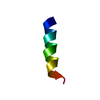
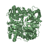
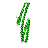
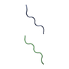
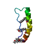
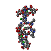
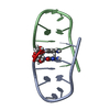
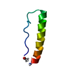
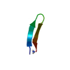
Entry Database : PDB / ID : 2y3lTitle Structure of segment MVGGVVIA from the amyloid-beta peptide (Ab, residues 35-42), alternate polymorph 2 AMYLOID BETA A4 PROTEIN Keywords / Function / homology Function Domain/homology Component
/ / / / / / / / / / / / / / / / / / / / / / / / / / / / / / / / / / / / / / / / / / / / / / / / / / / / / / / / / / / / / / / / / / / / / / / / / / / / / / / / / / / / / / / / / / / / / / / / / / / / / / / / / / / / / / / / / / / / / / / / / / / / / / / / / / / / / / / / / / / / / / / / / / / Biological species HOMO SAPIENS (human)Method / / / Resolution : 2.1 Å Authors Colletier, J.P. / Laganowsky, A. / Sawaya, M.R. / Eisenberg, D. Journal : Proc.Natl.Acad.Sci.USA / Year : 2011Title : Molecular Basis for Amyloid-{Beta} Polymorphism.Authors : Colletier, J. / Laganowsky, A. / Landau, M. / Zhao, M. / Soriaga, A.B. / Goldschmidt, L. / Flot, D. / Cascio, D. / Sawaya, M.R. / Eisenberg, D. History Deposition Dec 21, 2010 Deposition site / Processing site Revision 1.0 Nov 2, 2011 Provider / Type Revision 1.1 May 8, 2024 Group / Database references / OtherCategory chem_comp_atom / chem_comp_bond ... chem_comp_atom / chem_comp_bond / database_2 / pdbx_database_status Item / _database_2.pdbx_database_accession / _pdbx_database_status.status_code_sf
Show all Show less
 Movie
Movie Controller

 Yorodumi
 Open data
 Basic information
 Components
 Keywords
 ALZHEIMER DISEASE
 Function and homology information
 Authors
 Citation
 Structure visualization
 Downloads & links
 Other downloads


 PDBj
PDBj


 Assembly
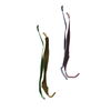
 Sample preparation
 / Beamline: 24-ID-E / Wavelength: 0.97918
  Processing