 Movie
Movie Controller


+ Open data

- Basic information
| Entry | Database: PDB / ID: 1ca0 | ||||||
|---|---|---|---|---|---|---|---|
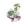
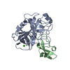
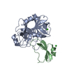
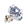
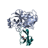
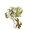
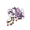
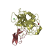
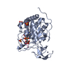
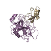
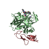
| Title | BOVINE CHYMOTRYPSIN COMPLEXED TO APPI | ||||||
 Components Components |
| ||||||
 Keywords Keywords | COMPLEX (SERINE PROTEASE/INHIBITOR) /  SERINE PROTEASE / INHIBITOR / PROTEASE-SUBSTRATE INTERACTIONS / COMPLEX (SERINE PROTEASE-INHIBITOR) / COMPLEX (SERINE PROTEASE-INHIBITOR) complex SERINE PROTEASE / INHIBITOR / PROTEASE-SUBSTRATE INTERACTIONS / COMPLEX (SERINE PROTEASE-INHIBITOR) / COMPLEX (SERINE PROTEASE-INHIBITOR) complex | ||||||
| Function / homology |  Function and homology informationchymotrypsin / regulation of epidermal growth factor-activated receptor activity / signaling receptor activator activity / collateral sprouting in absence of injury / cytosolic mRNA polyadenylation / microglia development / regulation of synapse structure or activity / Formyl peptide receptors bind formyl peptides and many other ligands / regulation of Wnt signaling pathway / axo-dendritic transport ...chymotrypsin / regulation of epidermal growth factor-activated receptor activity / signaling receptor activator activity / collateral sprouting in absence of injury / cytosolic mRNA polyadenylation / microglia development / regulation of synapse structure or activity / Formyl peptide receptors bind formyl peptides and many other ligands / regulation of Wnt signaling pathway / axo-dendritic transport / synaptic assembly at neuromuscular junction / smooth endoplasmic reticulum calcium ion homeostasis / axon midline choice point recognition / astrocyte activation involved in immune response / regulation of spontaneous synaptic transmission / mating behavior / NMDA selective glutamate receptor signaling pathway / ciliary rootlet / Lysosome Vesicle Biogenesis / PTB domain binding / Golgi-associated vesicle / positive regulation of amyloid fibril formation / neuron remodeling / Insertion of tail-anchored proteins into the endoplasmic reticulum membrane / protein serine/threonine kinase binding / Deregulated CDK5 triggers multiple neurodegenerative pathways in Alzheimer's disease models / : / nuclear envelope lumen / suckling behavior / presynaptic active zone / dendrite development / COPII-coated ER to Golgi transport vesicle / modulation of excitatory postsynaptic potential / TRAF6 mediated NF-kB activation / Advanced glycosylation endproduct receptor signaling / neuromuscular process controlling balance / The NLRP3 inflammasome / regulation of presynapse assembly / transition metal ion binding / regulation of multicellular organism growth / intracellular copper ion homeostasis / negative regulation of long-term synaptic potentiation / negative regulation of neuron differentiation / ECM proteoglycans / smooth endoplasmic reticulum / Mitochondrial protein degradation / positive regulation of T cell migration / spindle midzone / serpin family protein binding / Purinergic signaling in leishmaniasis infection / positive regulation of calcium-mediated signaling / forebrain development / clathrin-coated pit / regulation of peptidyl-tyrosine phosphorylation / positive regulation of chemokine production / Notch signaling pathway / serine protease inhibitor complex / digestion / positive regulation of G2/M transition of mitotic cell cycle / neuron projection maintenance / positive regulation of protein metabolic process / ionotropic glutamate receptor signaling pathway / positive regulation of glycolytic process / cholesterol metabolic process / response to interleukin-1 / positive regulation of mitotic cell cycle / extracellular matrix organization / axonogenesis / adult locomotory behavior / trans-Golgi network membrane / dendritic shaft / platelet alpha granule lumen / locomotory behavior / positive regulation of peptidyl-threonine phosphorylation / learning / positive regulation of interleukin-1 beta production / central nervous system development / positive regulation of long-term synaptic potentiation / endosome lumen / astrocyte activation / Post-translational protein phosphorylation / positive regulation of JNK cascade / synapse organization / regulation of long-term neuronal synaptic plasticity / microglial cell activation / TAK1-dependent IKK and NF-kappa-B activation / visual learning / serine-type endopeptidase inhibitor activity / neuromuscular junction / recycling endosome / cognition / positive regulation of inflammatory response / Golgi lumen / neuron cellular homeostasis / endocytosis / positive regulation of non-canonical NF-kappaB signal transduction / cellular response to amyloid-beta / positive regulation of interleukin-6 production / Regulation of Insulin-like Growth Factor (IGF) transport and uptake by Insulin-like Growth Factor Binding Proteins (IGFBPs) / neuron projection development Function and homology informationchymotrypsin / regulation of epidermal growth factor-activated receptor activity / signaling receptor activator activity / collateral sprouting in absence of injury / cytosolic mRNA polyadenylation / microglia development / regulation of synapse structure or activity / Formyl peptide receptors bind formyl peptides and many other ligands / regulation of Wnt signaling pathway / axo-dendritic transport ...chymotrypsin / regulation of epidermal growth factor-activated receptor activity / signaling receptor activator activity / collateral sprouting in absence of injury / cytosolic mRNA polyadenylation / microglia development / regulation of synapse structure or activity / Formyl peptide receptors bind formyl peptides and many other ligands / regulation of Wnt signaling pathway / axo-dendritic transport / synaptic assembly at neuromuscular junction / smooth endoplasmic reticulum calcium ion homeostasis / axon midline choice point recognition / astrocyte activation involved in immune response / regulation of spontaneous synaptic transmission / mating behavior / NMDA selective glutamate receptor signaling pathway / ciliary rootlet / Lysosome Vesicle Biogenesis / PTB domain binding / Golgi-associated vesicle / positive regulation of amyloid fibril formation / neuron remodeling / Insertion of tail-anchored proteins into the endoplasmic reticulum membrane / protein serine/threonine kinase binding / Deregulated CDK5 triggers multiple neurodegenerative pathways in Alzheimer's disease models / : / nuclear envelope lumen / suckling behavior / presynaptic active zone / dendrite development / COPII-coated ER to Golgi transport vesicle / modulation of excitatory postsynaptic potential / TRAF6 mediated NF-kB activation / Advanced glycosylation endproduct receptor signaling / neuromuscular process controlling balance / The NLRP3 inflammasome / regulation of presynapse assembly / transition metal ion binding / regulation of multicellular organism growth / intracellular copper ion homeostasis / negative regulation of long-term synaptic potentiation / negative regulation of neuron differentiation / ECM proteoglycans / smooth endoplasmic reticulum / Mitochondrial protein degradation / positive regulation of T cell migration / spindle midzone / serpin family protein binding / Purinergic signaling in leishmaniasis infection / positive regulation of calcium-mediated signaling / forebrain development / clathrin-coated pit / regulation of peptidyl-tyrosine phosphorylation / positive regulation of chemokine production / Notch signaling pathway / serine protease inhibitor complex / digestion / positive regulation of G2/M transition of mitotic cell cycle / neuron projection maintenance / positive regulation of protein metabolic process / ionotropic glutamate receptor signaling pathway / positive regulation of glycolytic process / cholesterol metabolic process / response to interleukin-1 / positive regulation of mitotic cell cycle / extracellular matrix organization / axonogenesis / adult locomotory behavior / trans-Golgi network membrane / dendritic shaft / platelet alpha granule lumen / locomotory behavior / positive regulation of peptidyl-threonine phosphorylation / learning / positive regulation of interleukin-1 beta production / central nervous system development / positive regulation of long-term synaptic potentiation / endosome lumen / astrocyte activation / Post-translational protein phosphorylation / positive regulation of JNK cascade / synapse organization / regulation of long-term neuronal synaptic plasticity / microglial cell activation / TAK1-dependent IKK and NF-kappa-B activation / visual learning / serine-type endopeptidase inhibitor activity / neuromuscular junction / recycling endosome / cognition / positive regulation of inflammatory response / Golgi lumen / neuron cellular homeostasis / endocytosis / positive regulation of non-canonical NF-kappaB signal transduction / cellular response to amyloid-beta / positive regulation of interleukin-6 production / Regulation of Insulin-like Growth Factor (IGF) transport and uptake by Insulin-like Growth Factor Binding Proteins (IGFBPs) / neuron projection developmentSimilarity search - Function | ||||||
| Biological species |  Bos taurus (cattle) Bos taurus (cattle) Homo sapiens (human) Homo sapiens (human) | ||||||
| Method | X-RAY DIFFRACTION / SYNCHROTRON / Resolution: 2.1 Å | ||||||
 Authors Authors | Scheidig, A.J. / Kossiakoff, A.A. | ||||||
 Citation Citation | Journal: Protein Sci. / Year: 1997 Title: Crystal structures of bovine chymotrypsin and trypsin complexed to the inhibitor domain of Alzheimer's amyloid beta-protein precursor (APPI) and basic pancreatic trypsin inhibitor (BPTI): ...Title: Crystal structures of bovine chymotrypsin and trypsin complexed to the inhibitor domain of Alzheimer's amyloid beta-protein precursor (APPI) and basic pancreatic trypsin inhibitor (BPTI): engineering of inhibitors with altered specificities. Authors: Scheidig, A.J. / Hynes, T.R. / Pelletier, L.A. / Wells, J.A. / Kossiakoff, A.A. #1: Journal: Biochemistry / Year: 1990Title: X-Ray Crystal Structure of the Protease Inhibitor Domain of Alzheimer'S Amyloid Beta-Protein Precursor Authors: Hynes, T.R. / Randal, M. / Kennedy, L.A. / Eigenbrot, C. / Kossiakoff, A.A. | ||||||
| History |
|
- Structure visualization
| Structure viewer | Molecule: MolmilJmol/JSmol |
|---|
- Downloads & links
-Download
| PDBx/mmCIF format | 1ca0.cif.gz | 119.4 KB | Display | PDBx/mmCIF format |
|---|---|---|---|---|
| PDB format | pdb1ca0.ent.gz | 99 KB | Display | PDB format |
| PDBx/mmJSON format | 1ca0.json.gz | Tree view | PDBx/mmJSON format | |
| Others |  Other downloads Other downloads |
-Validation report
| Arichive directory | https://data.pdbj.org/pub/pdb/validation_reports/ca/1ca0ftp://data.pdbj.org/pub/pdb/validation_reports/ca/1ca0 | HTTPS FTP |
|---|
-Related structure data

-Links
 PDBj
PDBj


- Assembly
| Deposited unit | 
| ||||||||
|---|---|---|---|---|---|---|---|---|---|
| 1 | 
| ||||||||
| 2 | 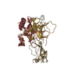
| ||||||||
| Unit cell |
| ||||||||
| Noncrystallographic symmetry (NCS) | NCS oper: (Code: given Matrix: (-0.782832, 0.018759, 0.62195), Vector : |
-Components
| #1: Protein/peptide | Mass: 1253.511 Da / Num. of mol.: 2 / Source method: isolated from a natural source / Source: (natural) Bos taurus (cattle) / References: UniProt: P00766, chymotrypsin#2: Protein | Mass: 13934.556 Da / Num. of mol.: 2 Source method: isolated from a genetically manipulated source Source: (gene. exp.) Bos taurus (cattle) / Gene: A4 / Organ: PANCREATICPancreas / Plasmid: PA4G32R / Gene (production host): A4 / Production host:  Escherichia coli (E. coli) / Strain (production host): 27C7 / References: UniProt: P00766, chymotrypsin Escherichia coli (E. coli) / Strain (production host): 27C7 / References: UniProt: P00766, chymotrypsin#3: Protein | Mass: 10074.495 Da / Num. of mol.: 2 Source method: isolated from a genetically manipulated source Source: (gene. exp.) Bos taurus (cattle) / Gene: A4 / Organ: PANCREATICPancreas / Plasmid: PA4G32R / Gene (production host): A4 / Production host: Escherichia coli (E. coli) / Strain (production host): 27C7 / References: UniProt: P00766, chymotrypsin#4: Protein | Mass: 6005.667 Da / Num. of mol.: 2 Source method: isolated from a genetically manipulated source Source: (gene. exp.) Homo sapiens (human) / Gene: A4 / Organ: PANCREATICPancreas / Plasmid: PA4G32R / Gene (production host): A4 / Production host: Escherichia coli (E. coli) / Strain (production host): 27C7 / References: UniProt: P05067#5: Water | ChemComp-HOH / | Water Mass: 18.015 Da / Num. of mol.: 262 / Source method: isolated from a natural source / Formula: H2O Mass: 18.015 Da / Num. of mol.: 262 / Source method: isolated from a natural source / Formula: H2O |
|---|
-Experimental details
-Experiment
| Experiment | Method: X-RAY DIFFRACTION / Number of used crystals: 1 |
|---|
- Sample preparation
| Crystal | Density Matthews: 2.39 Å3/Da / Density % sol: 48.09 % | ||||||||||||||||||||
|---|---|---|---|---|---|---|---|---|---|---|---|---|---|---|---|---|---|---|---|---|---|
| Crystal grow | pH: 4.5 / Details: pH 4.5 | ||||||||||||||||||||
| Crystal grow | *PLUS Temperature: 4 ℃ / Method: vapor diffusion, hanging drop | ||||||||||||||||||||
| Components of the solutions | *PLUS
|
-Data collection
| Diffraction | Mean temperature: 100 K |
|---|---|
| Diffraction source | Source: SYNCHROTRON / Site: SSRL  / Beamline: BL7-1 / Wavelength: 1.08 / Beamline: BL7-1 / Wavelength: 1.08 |
| Detector | Type: MARRESEARCH / Detector: IMAGE PLATE / Date: May 1, 1994 / Details: MIRROR |
| Radiation | Monochromator: MIRROR / Monochromatic (M) / Laue (L): M / Scattering type: x-ray |
| Radiation wavelength | Wavelength: 1.08 Å / Relative weight: 1 |
| Reflection | Resolution: 2.1→10 Å / Num. obs: 33088 / % possible obs: 81.1 % / Redundancy: 3.05 % / Biso Wilson estimate: 37.8 Å2 / Rsym value: 0.0298 / Net I/σ(I): 39.87 |
| Reflection | *PLUS Rmerge(I) obs: 0.03 |
- Processing
| Software |
| ||||||||||||||||||||||||||||||||||||||||||||||||||||||||||||
|---|---|---|---|---|---|---|---|---|---|---|---|---|---|---|---|---|---|---|---|---|---|---|---|---|---|---|---|---|---|---|---|---|---|---|---|---|---|---|---|---|---|---|---|---|---|---|---|---|---|---|---|---|---|---|---|---|---|---|---|---|---|
| Refinement | Resolution: 2.1→8 Å / Data cutoff high absF: 10000000 / Data cutoff low absF: 0.001 / Isotropic thermal model: RESTRAINED / Cross valid method: YES / σ(F): 0
| ||||||||||||||||||||||||||||||||||||||||||||||||||||||||||||
| Displacement parameters | Biso mean: 39.3 Å2 | ||||||||||||||||||||||||||||||||||||||||||||||||||||||||||||
| Refine analyze |
| ||||||||||||||||||||||||||||||||||||||||||||||||||||||||||||
| Refinement step | Cycle: LAST / Resolution: 2.1→8 Å
| ||||||||||||||||||||||||||||||||||||||||||||||||||||||||||||
| Refine LS restraints |
| ||||||||||||||||||||||||||||||||||||||||||||||||||||||||||||
| LS refinement shell | Resolution: 2.1→2.17 Å / Total num. of bins used: 10
| ||||||||||||||||||||||||||||||||||||||||||||||||||||||||||||
| Xplor file |
| ||||||||||||||||||||||||||||||||||||||||||||||||||||||||||||
| Software | *PLUS Name: X-PLOR / Version: 3.1 / Classification: refinement | ||||||||||||||||||||||||||||||||||||||||||||||||||||||||||||
| Refinement | *PLUS | ||||||||||||||||||||||||||||||||||||||||||||||||||||||||||||
| Solvent computation | *PLUS | ||||||||||||||||||||||||||||||||||||||||||||||||||||||||||||
| Displacement parameters | *PLUS | ||||||||||||||||||||||||||||||||||||||||||||||||||||||||||||
| Refine LS restraints | *PLUS
|
