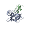
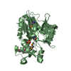
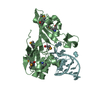
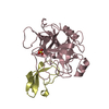
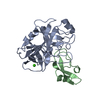
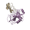
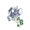
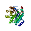
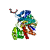
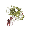
[English] 日本語
Yorodumi
- PDB-3l33: Human mesotrypsin complexed with amyloid precursor protein inhibi... -

+
Open data


ID or keywords:

Loading...

-
Basic information

Entry
Database: PDB / ID: 3l33
TitleHuman mesotrypsin complexed with amyloid precursor protein inhibitor(APPI)
Components
  • Amyloid beta A4 protein
  • Trypsin-3
KeywordsHydrolase/Cell Adhesion / Human mesotrypsin / alzheimer's amyloid precursor protein inhibitor / APPI / Serine protease inhibitor / Hydrolase-Cell Adhesion complex
Function / homology
Function and homology information


Uptake of dietary cobalamins into enterocytes / regulation of epidermal growth factor-activated receptor activity / signaling receptor activator activity / collateral sprouting in absence of injury / cytosolic mRNA polyadenylation / antimicrobial humoral response / microglia development / Alpha-defensins / regulation of synapse structure or activity / Formyl peptide receptors bind formyl peptides and many other ligands ...Uptake of dietary cobalamins into enterocytes / regulation of epidermal growth factor-activated receptor activity / signaling receptor activator activity / collateral sprouting in absence of injury / cytosolic mRNA polyadenylation / antimicrobial humoral response / microglia development / Alpha-defensins / regulation of synapse structure or activity / Formyl peptide receptors bind formyl peptides and many other ligands / regulation of Wnt signaling pathway / axo-dendritic transport / synaptic assembly at neuromuscular junction / smooth endoplasmic reticulum calcium ion homeostasis / axon midline choice point recognition / astrocyte activation involved in immune response / zymogen activation / regulation of spontaneous synaptic transmission / mating behavior / NMDA selective glutamate receptor signaling pathway / ciliary rootlet / Lysosome Vesicle Biogenesis / PTB domain binding / Antimicrobial peptides / Golgi-associated vesicle / positive regulation of amyloid fibril formation / neuron remodeling / Insertion of tail-anchored proteins into the endoplasmic reticulum membrane / protein serine/threonine kinase binding / Deregulated CDK5 triggers multiple neurodegenerative pathways in Alzheimer's disease models / : / nuclear envelope lumen / suckling behavior / modulation of excitatory postsynaptic potential / presynaptic active zone / dendrite development / COPII-coated ER to Golgi transport vesicle / TRAF6 mediated NF-kB activation / Advanced glycosylation endproduct receptor signaling / Mitochondrial protein degradation / neuromuscular process controlling balance / The NLRP3 inflammasome / regulation of presynapse assembly / transition metal ion binding / intracellular copper ion homeostasis / regulation of multicellular organism growth / negative regulation of long-term synaptic potentiation / negative regulation of neuron differentiation / ECM proteoglycans / smooth endoplasmic reticulum / endothelial cell migration / trypsin / positive regulation of T cell migration / spindle midzone / Purinergic signaling in leishmaniasis infection / positive regulation of calcium-mediated signaling / forebrain development / clathrin-coated pit / regulation of peptidyl-tyrosine phosphorylation / positive regulation of chemokine production / Notch signaling pathway / digestion / positive regulation of G2/M transition of mitotic cell cycle / positive regulation of protein metabolic process / ionotropic glutamate receptor signaling pathway / neuron projection maintenance / serine-type peptidase activity / cholesterol metabolic process / response to interleukin-1 / positive regulation of glycolytic process / positive regulation of mitotic cell cycle / extracellular matrix organization / axonogenesis / adult locomotory behavior / trans-Golgi network membrane / dendritic shaft / platelet alpha granule lumen / locomotory behavior / positive regulation of peptidyl-threonine phosphorylation / learning / positive regulation of interleukin-1 beta production / central nervous system development / positive regulation of long-term synaptic potentiation / endosome lumen / astrocyte activation / Post-translational protein phosphorylation / positive regulation of JNK cascade / synapse organization / regulation of long-term neuronal synaptic plasticity / microglial cell activation / TAK1-dependent IKK and NF-kappa-B activation / visual learning / serine-type endopeptidase inhibitor activity / neuromuscular junction / recycling endosome / cognition / positive regulation of inflammatory response / Golgi lumen / neuron cellular homeostasis / endocytosis
Similarity search - Function
Pancreatic trypsin inhibitor Kunitz domain / Factor Xa Inhibitor / Amyloidogenic glycoprotein, copper-binding / Amyloidogenic glycoprotein, copper-binding domain conserved site / Amyloidogenic glycoprotein, copper-binding domain superfamily / Copper-binding of amyloid precursor, CuBD / Amyloid precursor protein (APP) copper-binding (CuBD) domain signature. / Amyloidogenic glycoprotein, amyloid-beta peptide superfamily / Beta-amyloid peptide (beta-APP) / Amyloidogenic glycoprotein, amyloid-beta peptide ...Pancreatic trypsin inhibitor Kunitz domain / Factor Xa Inhibitor / Amyloidogenic glycoprotein, copper-binding / Amyloidogenic glycoprotein, copper-binding domain conserved site / Amyloidogenic glycoprotein, copper-binding domain superfamily / Copper-binding of amyloid precursor, CuBD / Amyloid precursor protein (APP) copper-binding (CuBD) domain signature. / Amyloidogenic glycoprotein, amyloid-beta peptide superfamily / Beta-amyloid peptide (beta-APP) / Amyloidogenic glycoprotein, amyloid-beta peptide / Beta-amyloid precursor protein C-terminal / Amyloidogenic glycoprotein, intracellular domain, conserved site / Beta-amyloid precursor protein C-terminus / Amyloid precursor protein (APP) intracellular domain signature. / Amyloid precursor protein (APP) E1 domain profile. / Amyloid precursor protein (APP) E2 domain profile. / Amyloidogenic glycoprotein, extracellular / Amyloidogenic glycoprotein, heparin-binding / Amyloidogenic glycoprotein, E2 domain / E2 domain superfamily / Amyloidogenic glycoprotein, heparin-binding domain superfamily / Amyloid A4 N-terminal heparin-binding / E2 domain of amyloid precursor protein / amyloid A4 / Amyloidogenic glycoprotein / Proteinase inhibitor I2, Kunitz, conserved site / Pancreatic trypsin inhibitor (Kunitz) family signature. / BPTI/Kunitz family of serine protease inhibitors. / Pancreatic trypsin inhibitor Kunitz domain / Kunitz/Bovine pancreatic trypsin inhibitor domain / Pancreatic trypsin inhibitor (Kunitz) family profile. / Pancreatic trypsin inhibitor Kunitz domain superfamily / Few Secondary Structures / Irregular / Serine proteases, trypsin family, histidine active site / Peptidase S1A, chymotrypsin family / Serine proteases, trypsin family, histidine active site. / Serine proteases, trypsin domain profile. / Trypsin-like serine protease / Serine proteases, trypsin domain / Trypsin / Trypsin-like serine proteases / PH-like domain superfamily / Thrombin, subunit H / Peptidase S1, PA clan, chymotrypsin-like fold / Peptidase S1, PA clan / Beta Barrel / Mainly Beta
Similarity search - Domain/homology
FORMIC ACID / Amyloid-beta precursor protein / Trypsin-3
Similarity search - Component
Biological speciesHomo sapiens (human)
MethodX-RAY DIFFRACTION / SYNCHROTRON / MOLECULAR REPLACEMENT / molecular replacement / Resolution: 2.48 Å
AuthorsSalameh, M.A. / Soares, A.S. / Radisky, E.S.
CitationJournal: J.Biol.Chem. / Year: 2010
Title: Determinants of affinity and proteolytic stability in interactions of Kunitz family protease inhibitors with mesotrypsin.
Authors: Salameh, M.A. / Soares, A.S. / Navaneetham, D. / Sinha, D. / Walsh, P.N. / Radisky, E.S.
History
DepositionDec 16, 2009Deposition site: RCSB / Processing site: RCSB
Revision 1.0Sep 22, 2010Provider: repository / Type: Initial release
Revision 1.1Jul 13, 2011Group: Version format compliance

-
Structure visualization

Structure viewerMolecule:
MolmilJmol/JSmol

Downloads & links

-
Assembly

Deposited unit
A: Trypsin-3
B: Trypsin-3
C: Trypsin-3
D: Trypsin-3
E: Amyloid beta A4 protein
F: Amyloid beta A4 protein
G: Amyloid beta A4 protein
H: Amyloid beta A4 protein
hetero molecules


Theoretical massNumber of molelcules
Total (without water)121,34331
Polymers120,3088
Non-polymers1,03523
Water4,125229
1
A: Trypsin-3
E: Amyloid beta A4 protein
hetero molecules


Theoretical massNumber of molelcules
Total (without water)30,53112
Polymers30,0772
Non-polymers45410
Water362
TypeNameSymmetry operationNumber
identity operation1_555x,y,z1
Buried area2450 Å2
ΔGint-23 kcal/mol
Surface area12000 Å2
MethodPISA
2
B: Trypsin-3
F: Amyloid beta A4 protein
hetero molecules


Theoretical massNumber of molelcules
Total (without water)30,3939
Polymers30,0772
Non-polymers3167
Water362
TypeNameSymmetry operationNumber
identity operation1_555x,y,z1
Buried area2290 Å2
ΔGint-22 kcal/mol
Surface area12170 Å2
MethodPISA
3
C: Trypsin-3
G: Amyloid beta A4 protein
hetero molecules


Theoretical massNumber of molelcules
Total (without water)30,2556
Polymers30,0772
Non-polymers1784
Water362
TypeNameSymmetry operationNumber
identity operation1_555x,y,z1
Buried area1780 Å2
ΔGint-20 kcal/mol
Surface area12090 Å2
MethodPISA
4
D: Trypsin-3
H: Amyloid beta A4 protein
hetero molecules


Theoretical massNumber of molelcules
Total (without water)30,1634
Polymers30,0772
Non-polymers862
Water362
TypeNameSymmetry operationNumber
identity operation1_555x,y,z1
Buried area1500 Å2
ΔGint-19 kcal/mol
Surface area12070 Å2
MethodPISA
Unit cell
Length a, b, c (Å)92.793, 130.067, 132.338
Angle α, β, γ (deg.)90.00, 90.00, 90.00
Int Tables number18
Space group name H-MP22121

-
Components

#1: Protein
Trypsin-3 / / Trypsin III / Brain trypsinogen / Mesotrypsinogen / Trypsin IV / Serine protease 3 / Serine protease 4


Mass: 24257.457 Da / Num. of mol.: 4 / Fragment: Trypsin-3 / Mutation: S195A
Source method: isolated from a genetically manipulated source
Source: (gene. exp.) Homo sapiens (human) / Strain: BL21(DE3) / Gene: PRSS3, PRSS4, TRY3, TRY4 / Production host: Escherichia coli (E. coli) / References: UniProt: P35030, trypsin
#2: Protein
Amyloid beta A4 protein / Alzheimer disease amyloid protein / ABPP / APPI / APP


Mass: 5819.501 Da / Num. of mol.: 4 / Fragment: UNP residues 290-341
Source method: isolated from a genetically manipulated source
Source: (gene. exp.) Homo sapiens (human) / Gene: APP, A4, AD1 / Production host: Pichia pastoris (fungus) / References: UniProt: P05067
#3: Chemical
ChemComp-FMT / FORMIC ACID / Formic acid


Mass: 46.025 Da / Num. of mol.: 19 / Source method: obtained synthetically / Formula: CH2O2
#4: Chemical
ChemComp-CA / CALCIUM ION


Mass: 40.078 Da / Num. of mol.: 4 / Source method: obtained synthetically / Formula: Ca
#5: Water ChemComp-HOH / water / Water


Mass: 18.015 Da / Num. of mol.: 229 / Source method: isolated from a natural source / Formula: H2O

-
Experimental details

-
Experiment

ExperimentMethod: X-RAY DIFFRACTION / Number of used crystals: 1

-
Sample preparation

CrystalDensity Matthews: 3.32 Å3/Da / Density % sol: 62.94 %
Crystal growTemperature: 298 K / Method: vapor diffusion, hanging drop
Details: 4M sodium formate solution, VAPOR DIFFUSION, HANGING DROP, temperature 298.0K

-
Data collection

Diffraction sourceSource: SYNCHROTRON / Site: NSLS / Beamline: X12B
RadiationProtocol: SINGLE WAVELENGTH / Monochromatic (M) / Laue (L): M / Scattering type: x-ray
Radiation wavelengthRelative weight: 1
ReflectionResolution: 2.48→23.702 Å / Num. all: 57050 / Num. obs: 57050 / % possible obs: 99.3 % / Redundancy: 14.2 % / Rmerge(I) obs: 0.263 / Rsym value: 0.263 / Net I/σ(I): 11.1
Reflection shellResolution: 2.48→2.61 Å / Redundancy: 12.4 % / Rmerge(I) obs: 0.024 / Mean I/σ(I) obs: 0.3 / Num. measured all: 98361 / Num. unique all: 7901 / Rsym value: 0.02353 / % possible all: 95.8

-
Phasing

PhasingMethod: molecular replacement

-
Processing

Software
NameVersionClassificationNB
MOSFLM3.2.25data reduction
SCALA3.2.25data scaling
PHASER2007_05_29_2026phasing
PHENIXrefinement
PDB_EXTRACT3.005data extraction
RefinementMethod to determine structure: MOLECULAR REPLACEMENT / Resolution: 2.48→23.702 Å / Occupancy max: 1 / Occupancy min: 0 / FOM work R set: 0.843 / Stereochemistry target values: mlhl
RfactorNum. reflection% reflection
Rfree0.256 1992 3.5 %
Rwork0.198 54882 -
obs-56874 99.08 %
Solvent computationBsol: 73.674 Å2 / ksol: 0.426 e/Å3
Displacement parametersBiso max: 222.85 Å2 / Biso mean: 44.887 Å2 / Biso min: 10.78 Å2
Baniso -1Baniso -2Baniso -3
1-7.24 Å2-0 Å2-0 Å2
2---6.667 Å20 Å2
3----0.572 Å2
Refinement stepCycle: LAST / Resolution: 2.48→23.702 Å
ProteinNucleic acidLigandSolventTotal
Num. atoms8412 0 61 229 8702
Refine LS restraints
Refine-IDTypeDev idealWeight
X-RAY DIFFRACTIONf_angle_d0.8711
X-RAY DIFFRACTIONf_bond_d0.0081
X-RAY DIFFRACTIONf_chiral_restr0.0631
X-RAY DIFFRACTIONf_dihedral_angle_d11.6661
X-RAY DIFFRACTIONf_plane_restr0.0031
X-RAY DIFFRACTIONf_nbd_refined4.0871
LS refinement shellResolution: 2.48→2.488 Å / Total num. of bins used: 110
RfactorNum. reflection% reflection
Rwork0.261 438 -
obs--82 %

+
About Yorodumi

-
News

-
Feb 9, 2022. New format data for meta-information of EMDB entries

New format data for meta-information of EMDB entries

  • Version 3 of the EMDB header file is now the official format.
  • The previous official version 1.9 will be removed from the archive.

Related info.:EMDB header

External links:wwPDB to switch to version 3 of the EMDB data model

-
Aug 12, 2020. Covid-19 info

Covid-19 info

URL: https://pdbj.org/emnavi/covid19.php

New page: Covid-19 featured information page in EM Navigator.

Related info.:Covid-19 info / Mar 5, 2020. Novel coronavirus structure data

+
Mar 5, 2020. Novel coronavirus structure data

Novel coronavirus structure data

Related info.:Yorodumi Speices / Aug 12, 2020. Covid-19 info

External links:COVID-19 featured content - PDBj / Molecule of the Month (242):Coronavirus Proteases

+
Jan 31, 2019. EMDB accession codes are about to change! (news from PDBe EMDB page)

EMDB accession codes are about to change! (news from PDBe EMDB page)

  • The allocation of 4 digits for EMDB accession codes will soon come to an end. Whilst these codes will remain in use, new EMDB accession codes will include an additional digit and will expand incrementally as the available range of codes is exhausted. The current 4-digit format prefixed with “EMD-” (i.e. EMD-XXXX) will advance to a 5-digit format (i.e. EMD-XXXXX), and so on. It is currently estimated that the 4-digit codes will be depleted around Spring 2019, at which point the 5-digit format will come into force.
  • The EM Navigator/Yorodumi systems omit the EMD- prefix.

Related info.:Q: What is EMD? / ID/Accession-code notation in Yorodumi/EM Navigator

External links:EMDB Accession Codes are Changing Soon! / Contact to PDBj

+
Jul 12, 2017. Major update of PDB

Major update of PDB

  • wwPDB released updated PDB data conforming to the new PDBx/mmCIF dictionary.
  • This is a major update changing the version number from 4 to 5, and with Remediation, in which all the entries are updated.
  • In this update, many items about electron microscopy experimental information are reorganized (e.g. em_software).
  • Now, EM Navigator and Yorodumi are based on the updated data.

External links:wwPDB Remediation / Enriched Model Files Conforming to OneDep Data Standards Now Available in the PDB FTP Archive

-
Yorodumi

Thousand views of thousand structures

  • Yorodumi is a browser for structure data from EMDB, PDB, SASBDB, etc.
  • This page is also the successor to EM Navigator detail page, and also detail information page/front-end page for Omokage search.
  • The word "yorodu" (or yorozu) is an old Japanese word meaning "ten thousand". "mi" (miru) is to see.

Related info.:EMDB / PDB / SASBDB / Comparison of 3 databanks / Yorodumi Search / Aug 31, 2016. New EM Navigator & Yorodumi / Yorodumi Papers / Jmol/JSmol / Function and homology information / Changes in new EM Navigator and Yorodumi

Read more