 Movie
Movie Controller

[English] 日本語
 Yorodumi
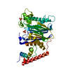
- PDB-2v64: Crystallographic structure of the conformational dimer of the Spi... -
+ Open data

- Basic information
| Entry | Database: PDB / ID: 2v64 | ||||||
|---|---|---|---|---|---|---|---|
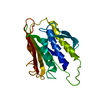
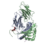
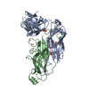
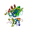
| Title | Crystallographic structure of the conformational dimer of the Spindle Assembly Checkpoint protein Mad2. | ||||||
 Components Components |
| ||||||
 Keywords Keywords |  CELL CYCLE / SPINDLE ASSEMBLY CHECKPOINT / MAD2 / NUCLEUS / MITOSIS / APOPTOSIS / CELL DIVISION / PHOSPHORYLATION / CONFORMATIONAL DIMER CELL CYCLE / SPINDLE ASSEMBLY CHECKPOINT / MAD2 / NUCLEUS / MITOSIS / APOPTOSIS / CELL DIVISION / PHOSPHORYLATION / CONFORMATIONAL DIMER | ||||||
| Function / homology |  Function and homology information Function and homology informationmitotic spindle assembly checkpoint MAD1-MAD2 complex / Inhibition of the proteolytic activity of APC/C required for the onset of anaphase by mitotic spindle checkpoint components / mitotic checkpoint complex / positive regulation of mitotic cell cycle spindle assembly checkpoint / establishment of centrosome localization / Inactivation of APC/C via direct inhibition of the APC/C complex / APC/C:Cdc20 mediated degradation of mitotic proteins / nuclear pore nuclear basket / negative regulation of ubiquitin protein ligase activity / mitotic spindle assembly checkpoint signaling ...mitotic spindle assembly checkpoint MAD1-MAD2 complex / Inhibition of the proteolytic activity of APC/C required for the onset of anaphase by mitotic spindle checkpoint components / mitotic checkpoint complex / positive regulation of mitotic cell cycle spindle assembly checkpoint / establishment of centrosome localization / Inactivation of APC/C via direct inhibition of the APC/C complex / APC/C:Cdc20 mediated degradation of mitotic proteins / nuclear pore nuclear basket / negative regulation of ubiquitin protein ligase activity / mitotic spindle assembly checkpoint signaling / mitotic sister chromatid segregation / establishment of mitotic spindle orientation / negative regulation of mitotic cell cycle / Amplification of signal from unattached kinetochores via a MAD2 inhibitory signal / Mitotic Prometaphase / EML4 and NUDC in mitotic spindle formation / Resolution of Sister Chromatid Cohesion / APC-Cdc20 mediated degradation of Nek2A / RHO GTPases Activate Formins / Cdc20:Phospho-APC/C mediated degradation of Cyclin A / negative regulation of protein catabolic process / mitotic spindle / kinetochore / spindle pole / Separation of Sister Chromatids / cell division / negative regulation of apoptotic process / perinuclear region of cytoplasm / protein homodimerization activity / nucleoplasm / identical protein binding / nucleus / cytosol / cytoplasmSimilarity search - Function | ||||||
| Biological species |  HOMO SAPIENS (human) HOMO SAPIENS (human)SYNTHETIC CONSTRUCT (others) | ||||||
| Method | X-RAY DIFFRACTION / SYNCHROTRON / MOLECULAR REPLACEMENT / Resolution: 2.9 Å | ||||||
 Authors Authors | Mapelli, M. / Massimiliano, L. / Santaguida, S. / Musacchio, A. | ||||||
 Citation Citation | Journal: Cell(Cambridge,Mass.) / Year: 2007 Title: The MAD2 Conformational Dimer: Structure and Implications for the Spindle Assembly Checkpoint Authors: Mapelli, M. / Massimiliano, L. / Santaguida, S. / Musacchio, A. | ||||||
| History |
|
- Structure visualization
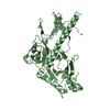
| Structure viewer | Molecule: MolmilJmol/JSmol |
|---|
- Downloads & links
-Download
| PDBx/mmCIF format | 2v64.cif.gz | 246.5 KB | Display | PDBx/mmCIF format |
|---|---|---|---|---|
| PDB format | pdb2v64.ent.gz | 198.7 KB | Display | PDB format |
| PDBx/mmJSON format | 2v64.json.gz | Tree view | PDBx/mmJSON format | |
| Others |  Other downloads Other downloads |
-Validation report
| Arichive directory | https://data.pdbj.org/pub/pdb/validation_reports/v6/2v64ftp://data.pdbj.org/pub/pdb/validation_reports/v6/2v64 | HTTPS FTP |
|---|
-Related structure data
| Related structure data |  1go4S S: Starting model for refinement |
|---|---|
| Similar structure data |


-Links
 PDBj
PDBj


- Assembly
| Deposited unit | 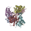
| ||||||||||||
|---|---|---|---|---|---|---|---|---|---|---|---|---|---|
| 1 | 
| ||||||||||||
| 2 | 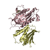
| ||||||||||||
| 3 | 
| ||||||||||||
| Unit cell |
| ||||||||||||
| Noncrystallographic symmetry (NCS) | NCS oper:
|
-Components
| #1: Protein | Mass: 24506.896 Da / Num. of mol.: 3 / Fragment: RESIDUES 2-205 Source method: isolated from a genetically manipulated source Source: (gene. exp.) HOMO SAPIENS (human) / Production host:  ESCHERICHIA COLI (E. coli) / Strain (production host): BL21 / References: UniProt: Q13257 ESCHERICHIA COLI (E. coli) / Strain (production host): BL21 / References: UniProt: Q13257#2: Protein/peptide | Alpha-enolaseMass: 1451.604 Da / Num. of mol.: 3 / Source method: obtained synthetically / Details: SEQUENCE SWYSYPPPQRAV / Source: (synth.) SYNTHETIC CONSTRUCT (others) #3: Protein | Mass: 23764.076 Da / Num. of mol.: 3 / Fragment: RESIDUES 2-108,118-205 Source method: isolated from a genetically manipulated source Source: (gene. exp.) HOMO SAPIENS (human) / Production host: ESCHERICHIA COLI (E. coli) / Strain (production host): BL21 / References: UniProt: Q13257#4: Water | ChemComp-HOH / | Water Mass: 18.015 Da / Num. of mol.: 6 / Source method: isolated from a natural source / Formula: H2O Mass: 18.015 Da / Num. of mol.: 6 / Source method: isolated from a natural source / Formula: H2OSequence details | N-TERMINAL HISTIDINE TAG IN CHAINS A, C, F. N-TERMINAL HISTIDINE TAG AND SUBSTITUTION OF RESIDUES ...N-TERMINAL HISTIDINE TAG IN CHAINS A, C, F. N-TERMINAL HISTIDINE TAG AND SUBSTITUTI | |
|---|
-Experimental details
-Experiment
| Experiment | Method: X-RAY DIFFRACTION / Number of used crystals: 1 |
|---|
- Sample preparation
| Crystal | Density Matthews: 2.8 Å3/Da / Density % sol: 55 % / Description: NONE |
|---|---|
| Crystal grow | pH: 4.6 / Details: 0.1M NAACETATE PH 4.6, 3.5M NAFORMATE |
-Data collection
| Diffraction | Mean temperature: 287 K |
|---|---|
| Diffraction source | Source: SYNCHROTRON / Site: ESRF  / Beamline: ID14-2 / Wavelength: 0.9333 / Beamline: ID14-2 / Wavelength: 0.9333 |
| Detector | Type: ADSC CCD / Detector: CCD / Date: Oct 10, 2006 |
| Radiation | Protocol: SINGLE WAVELENGTH / Monochromatic (M) / Laue (L): M / Scattering type: x-ray |
| Radiation wavelength | Wavelength: 0.9333 Å / Relative weight: 1 |
| Reflection | Resolution: 2.9→30 Å / Num. obs: 37591 / % possible obs: 100 % / Observed criterion σ(I): 3 / Redundancy: 6.9 % / Biso Wilson estimate: 33 Å2 / Rmerge(I) obs: 0.01 / Net I/σ(I): 16.2 |
| Reflection shell | Resolution: 2.9→3 Å / Redundancy: 6.9 % / Rmerge(I) obs: 0.45 / Mean I/σ(I) obs: 4.3 / % possible all: 100 |
- Processing
| Software |
| ||||||||||||||||||||||||||||||||||||||||||||||||||||||||||||
|---|---|---|---|---|---|---|---|---|---|---|---|---|---|---|---|---|---|---|---|---|---|---|---|---|---|---|---|---|---|---|---|---|---|---|---|---|---|---|---|---|---|---|---|---|---|---|---|---|---|---|---|---|---|---|---|---|---|---|---|---|---|
| Refinement | Method to determine structure: MOLECULAR REPLACEMENT Starting model: PDB ENTRY 1GO4 Resolution: 2.9→29.44 Å / Rfactor Rfree error: 0.007 / Data cutoff high absF: 2539666.54 / Isotropic thermal model: GROUP / Cross valid method: THROUGHOUT / σ(F): 0 / Stereochemistry target values: MAXIMUM LIKELIHOOD
| ||||||||||||||||||||||||||||||||||||||||||||||||||||||||||||
| Solvent computation | Solvent model: FLAT MODEL / Bsol: 33.5759 Å2 / ksol: 0.385076 e/Å3 | ||||||||||||||||||||||||||||||||||||||||||||||||||||||||||||
| Displacement parameters | Biso mean: 39.1 Å2
| ||||||||||||||||||||||||||||||||||||||||||||||||||||||||||||
| Refine analyze |
| ||||||||||||||||||||||||||||||||||||||||||||||||||||||||||||
| Refinement step | Cycle: LAST / Resolution: 2.9→29.44 Å
| ||||||||||||||||||||||||||||||||||||||||||||||||||||||||||||
| Refine LS restraints |
| ||||||||||||||||||||||||||||||||||||||||||||||||||||||||||||
| LS refinement shell | Resolution: 2.9→3.08 Å / Rfactor Rfree error: 0.021 / Total num. of bins used: 6
| ||||||||||||||||||||||||||||||||||||||||||||||||||||||||||||
| Xplor file |
|
