[English] 日本語
Yorodumi
- PDB-2uym: Crystal structure of KSP in complex with ADP and thiophene contai... -

+
Open data


ID or keywords:

Loading...

-
Basic information

Entry
Database: PDB / ID: 2uym
TitleCrystal structure of KSP in complex with ADP and thiophene containing inhibitor 37
ComponentsKINESIN-LIKE PROTEIN KIF11
KeywordsCELL CYCLE / KINESIN SPINDLE PROTEIN / KSP / EG5 / COMPLEX / INHIBITOR / THIOPHENE / ATP-BINDING / CELL DIVISION / COILED COIL / MICROTUBULE / MITOSIS / MOTOR PROTEIN / NUCLEOTIDE-BINDING / PHOSPHORYLATION
Function / homology
Function and homology information


spindle elongation / Kinesins / plus-end-directed microtubule motor activity / regulation of mitotic centrosome separation / mitotic centrosome separation / COPI-dependent Golgi-to-ER retrograde traffic / kinesin complex / microtubule motor activity / spindle organization / microtubule-based movement ...spindle elongation / Kinesins / plus-end-directed microtubule motor activity / regulation of mitotic centrosome separation / mitotic centrosome separation / COPI-dependent Golgi-to-ER retrograde traffic / kinesin complex / microtubule motor activity / spindle organization / microtubule-based movement / mitotic spindle assembly / MHC class II antigen presentation / mitotic spindle organization / mitotic spindle / spindle / spindle pole / mitotic cell cycle / microtubule binding / microtubule / cell division / protein kinase binding / protein-containing complex / ATP binding / membrane / nucleus / cytosol
Similarity search - Function
Kinesin-associated microtubule-binding domain / Kinesin-associated microtubule-binding / : / : / Kinesin motor domain / Kinesin / Kinesin motor domain signature. / Kinesin motor domain, conserved site / Kinesin motor domain / Kinesin motor domain profile. ...Kinesin-associated microtubule-binding domain / Kinesin-associated microtubule-binding / : / : / Kinesin motor domain / Kinesin / Kinesin motor domain signature. / Kinesin motor domain, conserved site / Kinesin motor domain / Kinesin motor domain profile. / Kinesin motor, catalytic domain. ATPase. / Kinesin motor domain / Kinesin motor domain superfamily / P-loop containing nucleoside triphosphate hydrolase / 3-Layer(aba) Sandwich / Alpha Beta
Similarity search - Domain/homology
ADENOSINE-5'-DIPHOSPHATE / Chem-K03 / Kinesin-like protein KIF11
Similarity search - Component
Biological speciesHOMO SAPIENS (human)
MethodX-RAY DIFFRACTION / SYNCHROTRON / MOLECULAR REPLACEMENT / Resolution: 2.11 Å
AuthorsLee, T.T.
CitationJournal: Bioorg.Med.Chem.Lett. / Year: 2007
Title: Synthesis and Sar of Thiophene Containing Kinesin Spindle Protein (Ksp) Inhibitors.
Authors: Pinkerton, A.B. / Lee, T.T. / Hoffman, T.Z. / Wang, Y. / Kahraman, M. / Cook, T.G. / Severance, D. / Gahman, T.C. / Noble, S.A. / Shiau, A.K. / Davis, R.L.
History
DepositionApr 10, 2007Deposition site: PDBE / Processing site: PDBE
Revision 1.0May 22, 2007Provider: repository / Type: Initial release
Revision 1.1May 8, 2011Group: Version format compliance
Revision 1.2Jul 13, 2011Group: Version format compliance
Revision 1.3Dec 13, 2023Group: Data collection / Database references ...Data collection / Database references / Derived calculations / Other / Refinement description
Category: chem_comp_atom / chem_comp_bond ...chem_comp_atom / chem_comp_bond / database_2 / pdbx_database_status / pdbx_initial_refinement_model / pdbx_struct_conn_angle / struct_conn / struct_site
Item: _database_2.pdbx_DOI / _database_2.pdbx_database_accession ..._database_2.pdbx_DOI / _database_2.pdbx_database_accession / _pdbx_database_status.status_code_sf / _pdbx_struct_conn_angle.ptnr1_auth_comp_id / _pdbx_struct_conn_angle.ptnr1_auth_seq_id / _pdbx_struct_conn_angle.ptnr1_label_asym_id / _pdbx_struct_conn_angle.ptnr1_label_atom_id / _pdbx_struct_conn_angle.ptnr1_label_comp_id / _pdbx_struct_conn_angle.ptnr1_label_seq_id / _pdbx_struct_conn_angle.ptnr3_auth_comp_id / _pdbx_struct_conn_angle.ptnr3_auth_seq_id / _pdbx_struct_conn_angle.ptnr3_label_asym_id / _pdbx_struct_conn_angle.ptnr3_label_atom_id / _pdbx_struct_conn_angle.ptnr3_label_comp_id / _pdbx_struct_conn_angle.ptnr3_label_seq_id / _pdbx_struct_conn_angle.value / _struct_conn.pdbx_dist_value / _struct_conn.ptnr1_auth_comp_id / _struct_conn.ptnr1_auth_seq_id / _struct_conn.ptnr1_label_asym_id / _struct_conn.ptnr1_label_atom_id / _struct_conn.ptnr1_label_comp_id / _struct_conn.ptnr1_label_seq_id / _struct_conn.ptnr2_auth_comp_id / _struct_conn.ptnr2_auth_seq_id / _struct_conn.ptnr2_label_asym_id / _struct_conn.ptnr2_label_atom_id / _struct_conn.ptnr2_label_comp_id / _struct_conn.ptnr2_label_seq_id / _struct_site.pdbx_auth_asym_id / _struct_site.pdbx_auth_comp_id / _struct_site.pdbx_auth_seq_id
Remark 700 
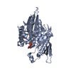
SHEET THE SHEET STRUCTURE OF THIS MOLECULE IS BIFURCATED. IN ORDER TO REPRESENT THIS FEATURE IN ... SHEET THE SHEET STRUCTURE OF THIS MOLECULE IS BIFURCATED. IN ORDER TO REPRESENT THIS FEATURE IN THE SHEET RECORDS BELOW, TWO SHEETS ARE DEFINED.
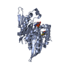
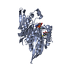
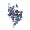
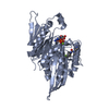
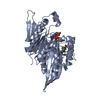
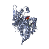
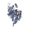
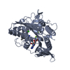
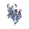
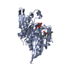
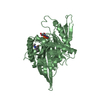
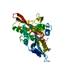
-
Structure visualization

Structure viewerMolecule:
MolmilJmol/JSmol

Downloads & links

-
Assembly

Deposited unit
A: KINESIN-LIKE PROTEIN KIF11
B: KINESIN-LIKE PROTEIN KIF11
hetero molecules


Theoretical massNumber of molelcules
Total (without water)83,7958
Polymers82,1112
Non-polymers1,6846
Water6,900383
1
A: KINESIN-LIKE PROTEIN KIF11
hetero molecules


Theoretical massNumber of molelcules
Total (without water)41,8984
Polymers41,0561
Non-polymers8423
Water181
TypeNameSymmetry operationNumber
identity operation1_555x,y,z1
MethodPQS
2
B: KINESIN-LIKE PROTEIN KIF11
hetero molecules


Theoretical massNumber of molelcules
Total (without water)41,8984
Polymers41,0561
Non-polymers8423
Water181
TypeNameSymmetry operationNumber
identity operation1_555x,y,z1
MethodPQS
Unit cell
Length a, b, c (Å)160.790, 80.452, 68.831
Angle α, β, γ (deg.)90.00, 96.16, 90.00
Int Tables number5
Space group name H-MC121
Components on special symmetry positions
IDModelComponents
11A-2064-

HOH

-
Components

#1: Protein KINESIN-LIKE PROTEIN KIF11 / / KINESIN SPINDLE PROTEIN / KINESIN-RELATED MOTOR PROTEIN EG5 / KINESIN-LIKE SPINDLE PROTEIN HKSP / ...KINESIN SPINDLE PROTEIN / KINESIN-RELATED MOTOR PROTEIN EG5 / KINESIN-LIKE SPINDLE PROTEIN HKSP / THYROID RECEPTOR-INTERACTING PROTEIN 5 / TRIP-5 / KINESIN-LIKE PROTEIN 1


Mass: 41055.582 Da / Num. of mol.: 2 / Fragment: CATALYTIC DOMAIN, RESIDUES 1-368
Source method: isolated from a genetically manipulated source
Source: (gene. exp.) HOMO SAPIENS (human) / Production host: ESCHERICHIA COLI (E. coli) / Strain (production host): BL21(DE3) / References: UniProt: P52732
#2: Chemical ChemComp-ADP / ADENOSINE-5'-DIPHOSPHATE / Adenosine diphosphate


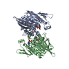
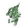
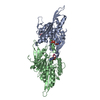
Mass: 427.201 Da / Num. of mol.: 2 / Source method: obtained synthetically / Formula: C10H15N5O10P2 / Comment: ADP, energy-carrying molecule*YM
#3: Chemical ChemComp-MG / MAGNESIUM ION


Mass: 24.305 Da / Num. of mol.: 2 / Source method: obtained synthetically / Formula: Mg
#4: Chemical ChemComp-K03 / N,N-DIETHYL-5,5-DIMETHYL-2-[(2-THIENYLCARBONYL)AMINO]-4,5,6,7-TETRAHYDRO-1-BENZOTHIOPHENE-3-CARBOXAMIDE


Mass: 390.563 Da / Num. of mol.: 2 / Source method: obtained synthetically / Formula: C20H26N2O2S2
#5: Water ChemComp-HOH / water / Water


Mass: 18.015 Da / Num. of mol.: 383 / Source method: isolated from a natural source / Formula: H2O
Compound detailsMOTOR PROTEIN NECESSARY FOR ESTABLISHING A BIPOLAR SPINDLE

-
Experimental details

-
Experiment

ExperimentMethod: X-RAY DIFFRACTION / Number of used crystals: 1

-
Sample preparation

CrystalDensity Matthews: 2.7 Å3/Da / Density % sol: 54 %
Crystal growpH: 6
Details: 100 MM BIS-TRIS (PH 6.0), 200 MM AMMONIUM SULFATE AND 18-20% PEG3350. 10 MM SRCL2

-
Data collection

DiffractionMean temperature: 277 K
Diffraction sourceSource: SYNCHROTRON / Site: ALS / Beamline: 5.0.2 / Wavelength: 1
DetectorType: ADSC CCD / Detector: CCD / Date: Sep 10, 2006
RadiationProtocol: SINGLE WAVELENGTH / Monochromatic (M) / Laue (L): M / Scattering type: x-ray
Radiation wavelengthWavelength: 1 Å / Relative weight: 1
ReflectionResolution: 2.1→50 Å / Num. obs: 49950 / % possible obs: 99.6 % / Observed criterion σ(I): 1.8 / Redundancy: 3.7 % / Rmerge(I) obs: 0.1 / Net I/σ(I): 13
Reflection shellResolution: 2.1→2.14 Å / Redundancy: 3.4 % / Rmerge(I) obs: 0.61 / Mean I/σ(I) obs: 1.8 / % possible all: 96.2

-
Processing

Software
NameVersionClassification
REFMAC5.2.0019refinement
DENZOdata reduction
SCALEPACKdata scaling
PHASERphasing
RefinementMethod to determine structure: MOLECULAR REPLACEMENT
Starting model: PDB ENTRY 1Q0B
Resolution: 2.11→49.45 Å / Cor.coef. Fo:Fc: 0.935 / Cor.coef. Fo:Fc free: 0.899 / SU B: 5.656 / SU ML: 0.153 / Cross valid method: THROUGHOUT / ESU R: 0.225 / ESU R Free: 0.205 / Stereochemistry target values: MAXIMUM LIKELIHOOD / Details: HYDROGENS HAVE BEEN ADDED IN THE RIDING POSITIONS.
RfactorNum. reflection% reflectionSelection details
Rfree0.274 2559 5.1 %RANDOM
Rwork0.217 ---
obs0.22 47209 98.8 %-
Solvent computationIon probe radii: 0.8 Å / Shrinkage radii: 0.8 Å / VDW probe radii: 1.4 Å / Solvent model: MASK
Displacement parametersBiso mean: 31.47 Å2
Baniso -1Baniso -2Baniso -3
1-0.47 Å20 Å2-1.77 Å2
2---1.54 Å20 Å2
3---0.7 Å2
Refinement stepCycle: LAST / Resolution: 2.11→49.45 Å
ProteinNucleic acidLigandSolventTotal
Num. atoms5198 0 108 383 5689
Refine LS restraints
Refine-IDTypeDev idealDev ideal targetNumber
X-RAY DIFFRACTIONr_bond_refined_d0.0120.0225388
X-RAY DIFFRACTIONr_bond_other_d
X-RAY DIFFRACTIONr_angle_refined_deg1.5831.9957300
X-RAY DIFFRACTIONr_angle_other_deg
X-RAY DIFFRACTIONr_dihedral_angle_1_deg6.2275658
X-RAY DIFFRACTIONr_dihedral_angle_2_deg35.40924.017234
X-RAY DIFFRACTIONr_dihedral_angle_3_deg17.1915976
X-RAY DIFFRACTIONr_dihedral_angle_4_deg18.8731542
X-RAY DIFFRACTIONr_chiral_restr0.1050.2850
X-RAY DIFFRACTIONr_gen_planes_refined0.0040.023954
X-RAY DIFFRACTIONr_gen_planes_other
X-RAY DIFFRACTIONr_nbd_refined0.2030.22401
X-RAY DIFFRACTIONr_nbd_other
X-RAY DIFFRACTIONr_nbtor_refined0.2980.23600
X-RAY DIFFRACTIONr_nbtor_other
X-RAY DIFFRACTIONr_xyhbond_nbd_refined0.1520.2391
X-RAY DIFFRACTIONr_xyhbond_nbd_other
X-RAY DIFFRACTIONr_metal_ion_refined
X-RAY DIFFRACTIONr_metal_ion_other
X-RAY DIFFRACTIONr_symmetry_vdw_refined0.1760.253
X-RAY DIFFRACTIONr_symmetry_vdw_other
X-RAY DIFFRACTIONr_symmetry_hbond_refined0.260.213
X-RAY DIFFRACTIONr_symmetry_hbond_other
X-RAY DIFFRACTIONr_symmetry_metal_ion_refined
X-RAY DIFFRACTIONr_symmetry_metal_ion_other
X-RAY DIFFRACTIONr_mcbond_it0.8471.53422
X-RAY DIFFRACTIONr_mcbond_other
X-RAY DIFFRACTIONr_mcangle_it1.44225350
X-RAY DIFFRACTIONr_mcangle_other
X-RAY DIFFRACTIONr_scbond_it1.76232241
X-RAY DIFFRACTIONr_scbond_other
X-RAY DIFFRACTIONr_scangle_it2.7774.51950
X-RAY DIFFRACTIONr_scangle_other
X-RAY DIFFRACTIONr_long_range_B_refined
X-RAY DIFFRACTIONr_long_range_B_other
X-RAY DIFFRACTIONr_rigid_bond_restr
X-RAY DIFFRACTIONr_sphericity_free
X-RAY DIFFRACTIONr_sphericity_bonded
LS refinement shellResolution: 2.11→2.16 Å / Total num. of bins used: 20 /
RfactorNum. reflection
Rfree0.303 167
Rwork0.257 2998

+
About Yorodumi

-
News

-
Feb 9, 2022. New format data for meta-information of EMDB entries

New format data for meta-information of EMDB entries

  • Version 3 of the EMDB header file is now the official format.
  • The previous official version 1.9 will be removed from the archive.

Related info.:EMDB header

External links:wwPDB to switch to version 3 of the EMDB data model

-
Aug 12, 2020. Covid-19 info

Covid-19 info

URL: https://pdbj.org/emnavi/covid19.php

New page: Covid-19 featured information page in EM Navigator.

Related info.:Covid-19 info / Mar 5, 2020. Novel coronavirus structure data

+
Mar 5, 2020. Novel coronavirus structure data

Novel coronavirus structure data

Related info.:Yorodumi Speices / Aug 12, 2020. Covid-19 info

External links:COVID-19 featured content - PDBj / Molecule of the Month (242):Coronavirus Proteases

+
Jan 31, 2019. EMDB accession codes are about to change! (news from PDBe EMDB page)

EMDB accession codes are about to change! (news from PDBe EMDB page)

  • The allocation of 4 digits for EMDB accession codes will soon come to an end. Whilst these codes will remain in use, new EMDB accession codes will include an additional digit and will expand incrementally as the available range of codes is exhausted. The current 4-digit format prefixed with “EMD-” (i.e. EMD-XXXX) will advance to a 5-digit format (i.e. EMD-XXXXX), and so on. It is currently estimated that the 4-digit codes will be depleted around Spring 2019, at which point the 5-digit format will come into force.
  • The EM Navigator/Yorodumi systems omit the EMD- prefix.

Related info.:Q: What is EMD? / ID/Accession-code notation in Yorodumi/EM Navigator

External links:EMDB Accession Codes are Changing Soon! / Contact to PDBj

+
Jul 12, 2017. Major update of PDB

Major update of PDB

  • wwPDB released updated PDB data conforming to the new PDBx/mmCIF dictionary.
  • This is a major update changing the version number from 4 to 5, and with Remediation, in which all the entries are updated.
  • In this update, many items about electron microscopy experimental information are reorganized (e.g. em_software).
  • Now, EM Navigator and Yorodumi are based on the updated data.

External links:wwPDB Remediation / Enriched Model Files Conforming to OneDep Data Standards Now Available in the PDB FTP Archive

-
Yorodumi

Thousand views of thousand structures

  • Yorodumi is a browser for structure data from EMDB, PDB, SASBDB, etc.
  • This page is also the successor to EM Navigator detail page, and also detail information page/front-end page for Omokage search.
  • The word "yorodu" (or yorozu) is an old Japanese word meaning "ten thousand". "mi" (miru) is to see.

Related info.:EMDB / PDB / SASBDB / Comparison of 3 databanks / Yorodumi Search / Aug 31, 2016. New EM Navigator & Yorodumi / Yorodumi Papers / Jmol/JSmol / Function and homology information / Changes in new EM Navigator and Yorodumi

Read more