 Movie
Movie Controller

[English] 日本語
 Yorodumi
- PDB-1ii6: Crystal Structure of the Mitotic Kinesin Eg5 in Complex with Mg-ADP. -
+ Open data

- Basic information
| Entry | Database: PDB / ID: 1ii6 | ||||||
|---|---|---|---|---|---|---|---|
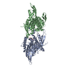
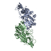
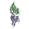
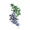
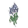
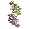
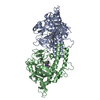
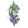
| Title | Crystal Structure of the Mitotic Kinesin Eg5 in Complex with Mg-ADP. | ||||||
 Components Components | KINESIN-RELATED MOTOR PROTEIN Eg5 | ||||||
 Keywords Keywords |  CELL CYCLE / Mg-ADP complex CELL CYCLE / Mg-ADP complex | ||||||
| Function / homology |  Function and homology information Function and homology informationspindle elongation / Kinesins / plus-end-directed microtubule motor activity / regulation of mitotic centrosome separation / mitotic centrosome separation / COPI-dependent Golgi-to-ER retrograde traffic / kinesin complex / microtubule motor activity / spindle organization / microtubule-based movement ...spindle elongation / Kinesins / plus-end-directed microtubule motor activity / regulation of mitotic centrosome separation / mitotic centrosome separation / COPI-dependent Golgi-to-ER retrograde traffic / kinesin complex / microtubule motor activity / spindle organization / microtubule-based movement / mitotic spindle assembly / MHC class II antigen presentation / mitotic spindle organization / mitotic spindle / spindle / spindle pole / mitotic cell cycle / microtubule binding / microtubule / cell division / protein kinase binding / protein-containing complex / ATP binding / membrane / nucleus / cytosolSimilarity search - Function | ||||||
| Biological species |  Homo sapiens (human) Homo sapiens (human) | ||||||
| Method | X-RAY DIFFRACTION / SYNCHROTRON / MOLECULAR REPLACEMENT / Resolution: 2.1 Å | ||||||
 Authors Authors | Turner, J. / Anderson, R. / Guo, J. / Beraud, C. / Sakowicz, R. / Fletterick, R. | ||||||
 Citation Citation | Journal: J.Biol.Chem. / Year: 2001 Title: Crystal structure of the mitotic spindle kinesin Eg5 reveals a novel conformation of the neck-linker. Authors: Turner, J. / Anderson, R. / Guo, J. / Beraud, C. / Fletterick, R. / Sakowicz, R. | ||||||
| History |
|
- Structure visualization
| Structure viewer | Molecule: MolmilJmol/JSmol |
|---|
- Downloads & links
-Download
| PDBx/mmCIF format | 1ii6.cif.gz | 152.9 KB | Display | PDBx/mmCIF format |
|---|---|---|---|---|
| PDB format | pdb1ii6.ent.gz | 120.4 KB | Display | PDB format |
| PDBx/mmJSON format | 1ii6.json.gz | Tree view | PDBx/mmJSON format | |
| Others |  Other downloads Other downloads |
-Validation report
| Arichive directory | https://data.pdbj.org/pub/pdb/validation_reports/ii/1ii6ftp://data.pdbj.org/pub/pdb/validation_reports/ii/1ii6 | HTTPS FTP |
|---|
-Related structure data
| Similar structure data |
|---|


-Links
 PDBj
PDBj


- Assembly
| Deposited unit | 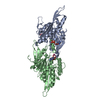
| ||||||||
|---|---|---|---|---|---|---|---|---|---|
| 1 |
| ||||||||
| Unit cell |
|
-Components
| #1: Protein | Mass: 41055.582 Da / Num. of mol.: 2 / Fragment: RESIDUES 1-368 Source method: isolated from a genetically manipulated source Source: (gene. exp.) Homo sapiens (human) / Gene: Eg5 / Plasmid: pET 43d / Species (production host): Escherichia coli / Production host:  Escherichia coli BL21(DE3) (bacteria) / Strain (production host): Bl21 (DE3) / References: UniProt: P52732 Escherichia coli BL21(DE3) (bacteria) / Strain (production host): Bl21 (DE3) / References: UniProt: P52732#2: Chemical | Nitrate  Mass: 62.005 Da / Num. of mol.: 2 / Source method: obtained synthetically / Formula: NO3 Mass: 62.005 Da / Num. of mol.: 2 / Source method: obtained synthetically / Formula: NO3#3: Chemical |   Mass: 24.305 Da / Num. of mol.: 2 / Source method: obtained synthetically / Formula: Mg Mass: 24.305 Da / Num. of mol.: 2 / Source method: obtained synthetically / Formula: Mg#4: Chemical | Adenosine diphosphate  Mass: 427.201 Da / Num. of mol.: 2 / Source method: obtained synthetically / Formula: C10H15N5O10P2 / Comment: ADP, energy-carrying molecule*YM Mass: 427.201 Da / Num. of mol.: 2 / Source method: obtained synthetically / Formula: C10H15N5O10P2 / Comment: ADP, energy-carrying molecule*YM#5: Water | ChemComp-HOH / | Water Mass: 18.015 Da / Num. of mol.: 351 / Source method: isolated from a natural source / Formula: H2O Mass: 18.015 Da / Num. of mol.: 351 / Source method: isolated from a natural source / Formula: H2O |
|---|
-Experimental details
-Experiment
| Experiment | Method: X-RAY DIFFRACTION / Number of used crystals: 3 |
|---|
- Sample preparation
| Crystal | Density Matthews: 2.39 Å3/Da / Density % sol: 48.44 % | ||||||||||||||||||||||||||||||
|---|---|---|---|---|---|---|---|---|---|---|---|---|---|---|---|---|---|---|---|---|---|---|---|---|---|---|---|---|---|---|---|
| Crystal grow | Temperature: 277 K / Method: vapor diffusion, sitting drop / pH: 5.6 Details: PEG 3350, MES, socium nitrate, pH 5.6, VAPOR DIFFUSION, SITTING DROP, temperature 277K | ||||||||||||||||||||||||||||||
| Crystal grow | *PLUS Temperature: 4 ℃ / pH: 6.8 / Details: used seeding | ||||||||||||||||||||||||||||||
| Components of the solutions | *PLUS
|
-Data collection
| Diffraction |
| ||||||||||||||||||||||||
|---|---|---|---|---|---|---|---|---|---|---|---|---|---|---|---|---|---|---|---|---|---|---|---|---|---|
| Diffraction source |
| ||||||||||||||||||||||||
| Detector |
| ||||||||||||||||||||||||
| Radiation |
| ||||||||||||||||||||||||
| Radiation wavelength | Wavelength: 0.97 Å / Relative weight: 1 | ||||||||||||||||||||||||
| Reflection | Resolution: 2.1→50 Å / Num. all: 236822 / Num. obs: 42896 / % possible obs: 94.9 % / Observed criterion σ(F): 2 / Observed criterion σ(I): 2 / Redundancy: 5.5 % / Rmerge(I) obs: 0.125 / Net I/σ(I): 9.4 | ||||||||||||||||||||||||
| Reflection shell | Resolution: 2.1→2.14 Å / Rmerge(I) obs: 0.103 / Num. unique all: 1924 / % possible all: 85 | ||||||||||||||||||||||||
| Reflection | *PLUS Num. measured all: 236822 | ||||||||||||||||||||||||
| Reflection shell | *PLUS % possible obs: 85 % |

- Processing
| Software |
| ||||||||||||||||||||||||||||||
|---|---|---|---|---|---|---|---|---|---|---|---|---|---|---|---|---|---|---|---|---|---|---|---|---|---|---|---|---|---|---|---|
| Refinement | Method to determine structure: MOLECULAR REPLACEMENT Starting model: catalytic core of KAR3 Resolution: 2.1→50 Å / Cross valid method: THROUGHOUT / σ(F): 2 / σ(I): 2
| ||||||||||||||||||||||||||||||
| Refinement step | Cycle: LAST / Resolution: 2.1→50 Å
| ||||||||||||||||||||||||||||||
| Software | *PLUS Name: CNS / Version: 1 / Classification: refinement | ||||||||||||||||||||||||||||||
| Refinement | *PLUS Highest resolution: 2.1 Å / Lowest resolution: 50 Å / σ(F): 2 / Rfactor obs: 0.2178 | ||||||||||||||||||||||||||||||
| Solvent computation | *PLUS | ||||||||||||||||||||||||||||||
| Displacement parameters | *PLUS | ||||||||||||||||||||||||||||||
| Refine LS restraints | *PLUS
|
