 Movie
Movie Controller


+ Open data

- Basic information
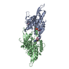
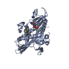
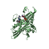
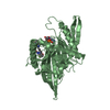
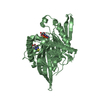
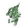
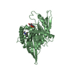
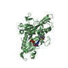
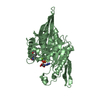
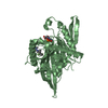
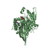
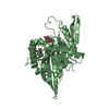
| Entry | Database: PDB / ID: 1x88 | ||||||
|---|---|---|---|---|---|---|---|
| Title | Human Eg5 motor domain bound to Mg-ADP and monastrol | ||||||
 Components Components | Kinesin-like protein KIF11 | ||||||
 Keywords Keywords | CELL CYCLE / Switch II / Motor domain / Neck linker | ||||||
| Function / homology |  Function and homology information Function and homology informationspindle elongation / Kinesins / plus-end-directed microtubule motor activity / regulation of mitotic centrosome separation / mitotic centrosome separation / COPI-dependent Golgi-to-ER retrograde traffic / kinesin complex / microtubule motor activity / spindle organization / microtubule-based movement ...spindle elongation / Kinesins / plus-end-directed microtubule motor activity / regulation of mitotic centrosome separation / mitotic centrosome separation / COPI-dependent Golgi-to-ER retrograde traffic / kinesin complex / microtubule motor activity / spindle organization / microtubule-based movement / mitotic spindle assembly / MHC class II antigen presentation / mitotic spindle organization / mitotic spindle / spindle / spindle pole / mitotic cell cycle / microtubule binding / microtubule / cell division / protein kinase binding / protein-containing complex / ATP binding / membrane / nucleus / cytosolSimilarity search - Function | ||||||
| Biological species |  Homo sapiens (human) Homo sapiens (human) | ||||||
| Method | X-RAY DIFFRACTION / SYNCHROTRON / MAD / Resolution: 1.8 Å | ||||||
 Authors Authors | Maliga, Z. / Mitchison, T.J. | ||||||
 Citation Citation | Journal: TO BE PUBLISHED Title: Structural Basis of Eg5 Inhibition by Monastrol Authors: Maliga, Z. / Mitchison, T.J. | ||||||
| History |
|
- Structure visualization
| Structure viewer | Molecule: MolmilJmol/JSmol |
|---|
- Downloads & links
-Download
| PDBx/mmCIF format | 1x88.cif.gz | 163.4 KB | Display | PDBx/mmCIF format |
|---|---|---|---|---|
| PDB format | pdb1x88.ent.gz | 126 KB | Display | PDB format |
| PDBx/mmJSON format | 1x88.json.gz | Tree view | PDBx/mmJSON format | |
| Others |  Other downloads Other downloads |
-Validation report
| Arichive directory | https://data.pdbj.org/pub/pdb/validation_reports/x8/1x88ftp://data.pdbj.org/pub/pdb/validation_reports/x8/1x88 | HTTPS FTP |
|---|
-Related structure data
| Related structure data | |
|---|---|
| Similar structure data |

-Links
 PDBj
PDBj


- Assembly
| Deposited unit | 
| ||||||||
|---|---|---|---|---|---|---|---|---|---|
| 1 | 
| ||||||||
| 2 | 
| ||||||||
| Unit cell |
|
-Components
| #1: Protein | / Kinesin-related motor protein Eg5 / Kinesin-like spindle protein HKSP / Thyroid receptor ...Kinesin-related motor protein Eg5 / Kinesin-like spindle protein HKSP / Thyroid receptor interacting protein 5 / TRIP5 / Kinesin-like protein 1 Mass: 40181.652 Da / Num. of mol.: 2 / Fragment: Kinesin-motor Source method: isolated from a genetically manipulated source Source: (gene. exp.) Homo sapiens (human) / Gene: KIF11, KNSL1, EG5 / Plasmid: pRSETa / Species (production host): Escherichia coli / Production host:  Escherichia coli BL21(DE3) (bacteria) / Strain (production host): BL21 (DE3) / References: UniProt: P52732 Escherichia coli BL21(DE3) (bacteria) / Strain (production host): BL21 (DE3) / References: UniProt: P52732#2: Chemical |   Mass: 24.305 Da / Num. of mol.: 2 / Source method: obtained synthetically / Formula: Mg Mass: 24.305 Da / Num. of mol.: 2 / Source method: obtained synthetically / Formula: Mg#3: Chemical | Adenosine diphosphate  Mass: 427.201 Da / Num. of mol.: 2 / Source method: obtained synthetically / Formula: C10H15N5O10P2 / Comment: ADP, energy-carrying molecule*YM Mass: 427.201 Da / Num. of mol.: 2 / Source method: obtained synthetically / Formula: C10H15N5O10P2 / Comment: ADP, energy-carrying molecule*YM#4: Chemical | Monastrol  Mass: 292.353 Da / Num. of mol.: 2 / Source method: obtained synthetically / Formula: C14H16N2O3S / Comment: inhibitor*YM Mass: 292.353 Da / Num. of mol.: 2 / Source method: obtained synthetically / Formula: C14H16N2O3S / Comment: inhibitor*YM#5: Water | ChemComp-HOH / | Water Mass: 18.015 Da / Num. of mol.: 698 / Source method: isolated from a natural source / Formula: H2O Mass: 18.015 Da / Num. of mol.: 698 / Source method: isolated from a natural source / Formula: H2O |
|---|
-Experimental details
-Experiment
| Experiment | Method: X-RAY DIFFRACTION / Number of used crystals: 2 |
|---|
- Sample preparation
| Crystal | Density Matthews: 2.75 Å3/Da / Density % sol: 55.32 % |
|---|---|
| Crystal grow | Temperature: 275 K / Method: vapor diffusion, hanging drop / pH: 6.9 Details: PEG3350, ammonium tartrate, potassium chloride, adenosine diphosphate, monastrol, sodium azide, PIPES, pH 6.9, VAPOR DIFFUSION, HANGING DROP, temperature 275K |
-Data collection
| Diffraction |
| ||||||||||||||||||
|---|---|---|---|---|---|---|---|---|---|---|---|---|---|---|---|---|---|---|---|
| Diffraction source |
| ||||||||||||||||||
| Detector |
| ||||||||||||||||||
| Radiation |
| ||||||||||||||||||
| Radiation wavelength |
| ||||||||||||||||||
| Reflection | Resolution: 1.7→20 Å / Num. all: 98099 / Num. obs: 93881 / % possible obs: 95.7 % / Observed criterion σ(F): 2 / Observed criterion σ(I): 2 / Redundancy: 10 % / Biso Wilson estimate: 13.4 Å2 | ||||||||||||||||||
| Reflection shell | Resolution: 1.7→1.76 Å / Redundancy: 4.4 % / % possible all: 73.6 |

- Processing
| Software |
| ||||||||||||||||||||||||||||||||||||||||||||||||||||||||||||||||||||||||||||||||
|---|---|---|---|---|---|---|---|---|---|---|---|---|---|---|---|---|---|---|---|---|---|---|---|---|---|---|---|---|---|---|---|---|---|---|---|---|---|---|---|---|---|---|---|---|---|---|---|---|---|---|---|---|---|---|---|---|---|---|---|---|---|---|---|---|---|---|---|---|---|---|---|---|---|---|---|---|---|---|---|---|---|
| Refinement | Method to determine structure: MAD Starting model: Chain building into electron density map obtained from Solve using MAD Resolution: 1.8→19.93 Å / Rfactor Rfree error: 0.003 / Data cutoff high absF: 291975.22 / Data cutoff low absF: 0 / Isotropic thermal model: RESTRAINED / Cross valid method: THROUGHOUT / σ(F): 0 / σ(I): 0 / Stereochemistry target values: Engh & Huber
| ||||||||||||||||||||||||||||||||||||||||||||||||||||||||||||||||||||||||||||||||
| Solvent computation | Solvent model: FLAT MODEL / Bsol: 42.0124 Å2 / ksol: 0.365705 e/Å3 | ||||||||||||||||||||||||||||||||||||||||||||||||||||||||||||||||||||||||||||||||
| Displacement parameters | Biso mean: 23.6 Å2
| ||||||||||||||||||||||||||||||||||||||||||||||||||||||||||||||||||||||||||||||||
| Refine analyze |
| ||||||||||||||||||||||||||||||||||||||||||||||||||||||||||||||||||||||||||||||||
| Refinement step | Cycle: LAST / Resolution: 1.8→19.93 Å
| ||||||||||||||||||||||||||||||||||||||||||||||||||||||||||||||||||||||||||||||||
| Refine LS restraints |
| ||||||||||||||||||||||||||||||||||||||||||||||||||||||||||||||||||||||||||||||||
| LS refinement shell | Resolution: 1.8→1.91 Å / Rfactor Rfree error: 0.007 / Total num. of bins used: 6
| ||||||||||||||||||||||||||||||||||||||||||||||||||||||||||||||||||||||||||||||||
| Xplor file |
|
