Type: MARMOSAIC 225 mm CCD / Detector: CCD / Date: Nov 15, 2006
Radiation
Protocol: SINGLE WAVELENGTH / Monochromatic (M) / Laue (L): M / Scattering type: x-ray
Radiation wavelength
Wavelength: 0.97903 Å / Relative weight: 1
Reflection
Redundancy: 15.9 % / Av σ(I) over netI: 15.1 / Number: 1496125 / Rmerge(I) obs: 0.097 / Χ2: 1.86 / D res high: 1.8 Å / D res low: 50 Å / Num. obs: 94229 / % possible obs: 99.5
Diffraction reflection shell
Highest resolution (Å)
Lowest resolution (Å)
% possible obs (%)
ID
Rmerge(I) obs
Chi squared
Redundancy
3.88
50
99.1
1
0.07
2.738
20.5
3.08
3.88
100
1
0.079
2.567
20.9
2.69
3.08
100
1
0.094
2.367
19.7
2.44
2.69
100
1
0.114
2.052
18.1
2.27
2.44
100
1
0.132
1.568
16.7
2.13
2.27
100
1
0.177
1.489
15.7
2.03
2.13
100
1
0.218
1.205
14.9
1.94
2.03
100
1
0.282
0.923
14
1.86
1.94
99.6
1
0.421
1.008
10.2
1.8
1.86
96.3
1
0.499
0.822
7.3
Reflection
Resolution: 1.8→50 Å / Num. obs: 94229 / % possible obs: 99.5 % / Redundancy: 15.9 % / Rsym value: 0.097 / Χ2: 1.857 / Net I/σ(I): 15.1
Reflection shell
Resolution (Å)
Redundancy (%)
Rmerge(I) obs
Num. unique all
Χ2
Diffraction-ID
% possible all
1.8-1.86
7.3
0.499
9032
0.822
1
96.3
1.86-1.94
10.2
0.421
9351
1.008
1
99.6
1.94-2.03
14
0.282
9393
0.923
1
100
2.03-2.13
14.9
0.218
9382
1.205
1
100
2.13-2.27
15.7
0.177
9407
1.489
1
100
2.27-2.44
16.7
0.132
9412
1.568
1
100
2.44-2.69
18.1
0.114
9462
2.052
1
100
2.69-3.08
19.7
0.094
9491
2.367
1
100
3.08-3.88
20.9
0.079
9541
2.567
1
100
3.88-50
20.5
0.07
9758
2.738
1
99.1
-
Processing
Software
Name
Version
Classification
NB
DENZO
datareduction
SCALEPACK
datascaling
REFMAC
5.2.0019
refinement
PDB_EXTRACT
2
dataextraction
SERGUI
datacollection
HKL-2000
datareduction
SOLVE
phasing
Refinement
Method to determine structure: SAD / Resolution: 1.8→30 Å / Cor.coef. Fo:Fc: 0.944 / Cor.coef. Fo:Fc free: 0.934 / SU B: 2.42 / SU ML: 0.076 / Cross valid method: THROUGHOUT / σ(F): 0 / ESU R: 0.123 / ESU R Free: 0.114 / Stereochemistry target values: MAXIMUM LIKELIHOOD / Details: HYDROGENS HAVE BEEN ADDED IN THE RIDING POSITIONS
Rfactor
Num. reflection
% reflection
Selection details
Rfree
0.226
4715
5 %
RANDOM
Rwork
0.205
-
-
-
all
0.206
94153
-
-
obs
0.206
94153
99.48 %
-
Solvent computation
Ion probe radii: 0.8 Å / Shrinkage radii: 0.8 Å / VDW probe radii: 1.2 Å / Solvent model: MASK
Displacement parameters
Biso mean: 20.529 Å2
Baniso -1
Baniso -2
Baniso -3
1-
0.5 Å2
0.25 Å2
0 Å2
2-
-
0.5 Å2
0 Å2
3-
-
-
-0.75 Å2
Refinement step
Cycle: LAST / Resolution: 1.8→30 Å
Protein
Nucleic acid
Ligand
Solvent
Total
Num. atoms
6032
0
106
566
6704
Refine LS restraints
Refine-ID
Type
Dev ideal
Dev ideal target
Number
X-RAY DIFFRACTION
r_bond_refined_d
0.009
0.022
6280
X-RAY DIFFRACTION
r_angle_refined_deg
1.111
1.97
8504
X-RAY DIFFRACTION
r_dihedral_angle_1_deg
4.873
5
755
X-RAY DIFFRACTION
r_dihedral_angle_2_deg
36.355
23.724
290
X-RAY DIFFRACTION
r_dihedral_angle_3_deg
13.19
15
1085
X-RAY DIFFRACTION
r_dihedral_angle_4_deg
14.162
15
42
X-RAY DIFFRACTION
r_chiral_restr
0.078
0.2
915
X-RAY DIFFRACTION
r_gen_planes_refined
0.004
0.02
4722
X-RAY DIFFRACTION
r_nbd_refined
0.197
0.2
3125
X-RAY DIFFRACTION
r_nbtor_refined
0.3
0.2
4328
X-RAY DIFFRACTION
r_xyhbond_nbd_refined
0.132
0.2
517
X-RAY DIFFRACTION
r_symmetry_vdw_refined
0.208
0.2
99
X-RAY DIFFRACTION
r_symmetry_hbond_refined
0.198
0.2
24
X-RAY DIFFRACTION
r_mcbond_it
0.656
1.5
3869
X-RAY DIFFRACTION
r_mcangle_it
0.963
2
6020
X-RAY DIFFRACTION
r_scbond_it
1.666
3
2915
X-RAY DIFFRACTION
r_scangle_it
2.668
4.5
2484
LS refinement shell
Resolution: 1.8→1.848 Å / Total num. of bins used: 20
Rfactor
Num. reflection
% reflection
Rfree
0.334
347
-
Rwork
0.273
6238
-
obs
-
6585
95.45 %
+
About Yorodumi
-
News
-
Feb 9, 2022. New format data for meta-information of EMDB entries
New format data for meta-information of EMDB entries
Version 3 of the EMDB header file is now the official format.
The previous official version 1.9 will be removed from the archive.
In the structure databanks used in Yorodumi, some data are registered as the other names, "COVID-19 virus" and "2019-nCoV". Here are the details of the virus and the list of structure data.
Jan 31, 2019. EMDB accession codes are about to change! (news from PDBe EMDB page)
EMDB accession codes are about to change! (news from PDBe EMDB page)
The allocation of 4 digits for EMDB accession codes will soon come to an end. Whilst these codes will remain in use, new EMDB accession codes will include an additional digit and will expand incrementally as the available range of codes is exhausted. The current 4-digit format prefixed with “EMD-” (i.e. EMD-XXXX) will advance to a 5-digit format (i.e. EMD-XXXXX), and so on. It is currently estimated that the 4-digit codes will be depleted around Spring 2019, at which point the 5-digit format will come into force.
The EM Navigator/Yorodumi systems omit the EMD- prefix.
Related info.:Q: What is EMD? / ID/Accession-code notation in Yorodumi/EM Navigator
Yorodumi is a browser for structure data from EMDB, PDB, SASBDB, etc.
This page is also the successor to EM Navigator detail page, and also detail information page/front-end page for Omokage search.
The word "yorodu" (or yorozu) is an old Japanese word meaning "ten thousand". "mi" (miru) is to see.
Related info.:EMDB / PDB / SASBDB / Comparison of 3 databanks / Yorodumi Search / Aug 31, 2016. New EM Navigator & Yorodumi / Yorodumi Papers / Jmol/JSmol / Function and homology information / Changes in new EM Navigator and Yorodumi
 Movie
Movie Controller

 Yorodumi
 Open data
 Basic information
 Components
 Keywords
 Function and homology information
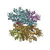
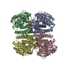
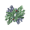
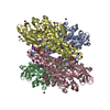
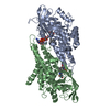
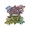
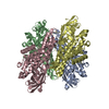
 Geobacillus kaustophilus (bacteria)
 Authors
 Citation
 Structure visualization
 Downloads & links
 Other downloads
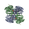


 PDBj
PDBj Assembly

 Mass: 785.550 Da / Num. of mol.: 2 / Source method: obtained synthetically / Formula: C27H33N9O15P2 / Comment: FAD*YM
 Mass: 18.015 Da / Num. of mol.: 566 / Source method: isolated from a natural source / Formula: H2O
 Sample preparation
 / Beamline: 22-BM / Wavelength: 0.97903 Å
 Processing