 Movie
Movie Controller

[English] 日本語
 Yorodumi
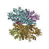
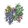
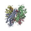
- PDB-3mdd: CRYSTAL STRUCTURES OF MEDIUM CHAIN ACYL-COA DEHYDROGENASE FROM PI... -
+ Open data

- Basic information
| Entry | Database: PDB / ID: 3mdd | ||||||
|---|---|---|---|---|---|---|---|
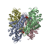
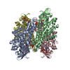
| Title | CRYSTAL STRUCTURES OF MEDIUM CHAIN ACYL-COA DEHYDROGENASE FROM PIG LIVER MITOCHONDRIA WITH AND WITHOUT SUBSTRATE | ||||||
 Components Components | MEDIUM CHAIN ACYL-COA DEHYDROGENASE | ||||||
 Keywords Keywords |  OXIDOREDUCTASE OXIDOREDUCTASE | ||||||
| Function / homology |  Function and homology information Function and homology informationmitochondrial fatty acid beta-oxidation of unsaturated fatty acids / Beta oxidation of decanoyl-CoA to octanoyl-CoA-CoA / Beta oxidation of octanoyl-CoA to hexanoyl-CoA / medium-chain fatty acid catabolic process / carnitine metabolic process, CoA-linked / medium-chain acyl-CoA dehydrogenase / medium-chain fatty acyl-CoA dehydrogenase activity / carnitine biosynthetic process / fatty acid beta-oxidation using acyl-CoA dehydrogenase / acyl-CoA dehydrogenase activity ...mitochondrial fatty acid beta-oxidation of unsaturated fatty acids / Beta oxidation of decanoyl-CoA to octanoyl-CoA-CoA / Beta oxidation of octanoyl-CoA to hexanoyl-CoA / medium-chain fatty acid catabolic process / carnitine metabolic process, CoA-linked / medium-chain acyl-CoA dehydrogenase / medium-chain fatty acyl-CoA dehydrogenase activity / carnitine biosynthetic process / fatty acid beta-oxidation using acyl-CoA dehydrogenase / acyl-CoA dehydrogenase activity / cardiac muscle cell differentiation / glycogen biosynthetic process / regulation of gluconeogenesis / fatty acid beta-oxidation / response to starvation / response to cold / post-embryonic development / liver development / mitochondrial membrane / flavin adenine dinucleotide binding / mitochondrial matrix / axon / mitochondrion / identical protein binding / cytoplasmSimilarity search - Function | ||||||
| Biological species |  Sus scrofa (pig) Sus scrofa (pig) | ||||||
| Method | X-RAY DIFFRACTION / Resolution: 2.4 Å | ||||||
 Authors Authors | Kim, J.-J.P. / Wang, M. / Paschke, R. | ||||||
 Citation Citation | Journal: Proc.Natl.Acad.Sci.USA / Year: 1993 Title: Crystal structures of medium-chain acyl-CoA dehydrogenase from pig liver mitochondria with and without substrate. Authors: Kim, J.J. / Wang, M. / Paschke, R. #1: Journal: Proc.Natl.Acad.Sci.USA / Year: 1988Title: Structure of the Medium-Chain Acyl-Coa Dehydrogenase from Pig Liver Mitochondria at 3-Angstroms Resolution Authors: Kim, J.-J.P. / Wu, J. | ||||||
| History |
| ||||||
| Remark 700 | SHEET STRAND 4 OF SHEET 2 DOES NOT INCLUDE RESIDUE 229. |
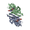
- Structure visualization
| Structure viewer | Molecule: MolmilJmol/JSmol |
|---|
- Downloads & links
-Download
| PDBx/mmCIF format | 3mdd.cif.gz | 160.3 KB | Display | PDBx/mmCIF format |
|---|---|---|---|---|
| PDB format | pdb3mdd.ent.gz | 126.5 KB | Display | PDB format |
| PDBx/mmJSON format | 3mdd.json.gz | Tree view | PDBx/mmJSON format | |
| Others |  Other downloads Other downloads |
-Validation report
| Arichive directory | https://data.pdbj.org/pub/pdb/validation_reports/md/3mddftp://data.pdbj.org/pub/pdb/validation_reports/md/3mdd | HTTPS FTP |
|---|
-Related structure data

-Links
 PDBj
PDBj- Assembly
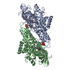
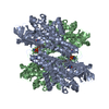
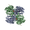
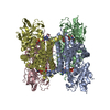
| Deposited unit | 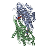
| ||||||||
|---|---|---|---|---|---|---|---|---|---|
| 1 | 
| ||||||||
| Unit cell |
|
-Components
| #1: Protein | Mass: 42419.289 Da / Num. of mol.: 2 Source method: isolated from a genetically manipulated source Source: (gene. exp.) Sus scrofa (pig) / References: UniProt: P41367, EC: 1.3.99.3#2: Chemical | Flavin adenine dinucleotide  Mass: 785.550 Da / Num. of mol.: 2 / Source method: obtained synthetically / Formula: C27H33N9O15P2 / Comment: FAD*YM Mass: 785.550 Da / Num. of mol.: 2 / Source method: obtained synthetically / Formula: C27H33N9O15P2 / Comment: FAD*YM#3: Water | ChemComp-HOH / | Water Mass: 18.015 Da / Num. of mol.: 166 / Source method: isolated from a natural source / Formula: H2O Mass: 18.015 Da / Num. of mol.: 166 / Source method: isolated from a natural source / Formula: H2O |
|---|
-Experimental details
-Experiment
| Experiment | Method: X-RAY DIFFRACTION |
|---|
- Sample preparation
| Crystal | Density Matthews: 2.74 Å3/Da / Density % sol: 55.13 % |
|---|---|
| Crystal grow | *PLUS Method: vapor diffusion, sitting drop |
-Data collection
| Reflection | *PLUS Highest resolution: 2.4 Å / Num. obs: 30930 / % possible obs: 84 % / Num. measured all: 138867 / Rmerge(I) obs: 0.047 |
|---|
- Processing
| Software | Name: GPRLSA / Classification: refinement | |||||||||||||||||||||||||||||||||||||||||||||||||||||||||||||||
|---|---|---|---|---|---|---|---|---|---|---|---|---|---|---|---|---|---|---|---|---|---|---|---|---|---|---|---|---|---|---|---|---|---|---|---|---|---|---|---|---|---|---|---|---|---|---|---|---|---|---|---|---|---|---|---|---|---|---|---|---|---|---|---|---|
| Refinement | Resolution: 2.4→6 Å / σ(F): 3 /
| |||||||||||||||||||||||||||||||||||||||||||||||||||||||||||||||
| Refinement step | Cycle: LAST / Resolution: 2.4→6 Å
| |||||||||||||||||||||||||||||||||||||||||||||||||||||||||||||||
| Refine LS restraints |
| |||||||||||||||||||||||||||||||||||||||||||||||||||||||||||||||
| Software | *PLUS Name: GPRLSA / Classification: refinement | |||||||||||||||||||||||||||||||||||||||||||||||||||||||||||||||
| Refinement | *PLUS Rfactor obs: 0.173 | |||||||||||||||||||||||||||||||||||||||||||||||||||||||||||||||
| Solvent computation | *PLUS | |||||||||||||||||||||||||||||||||||||||||||||||||||||||||||||||
| Displacement parameters | *PLUS Biso mean: 17.18 Å2 | |||||||||||||||||||||||||||||||||||||||||||||||||||||||||||||||
| Refine LS restraints | *PLUS Type: p_angle_d / Dev ideal: 0.021 |
