- PDB-1rx0: Crystal structure of isobutyryl-CoA dehydrogenase complexed with ... -
+
Open data
ID or keywords:
Loading...
-
Basic information
Entry
Database: PDB / ID: 1rx0
Title
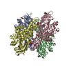
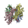
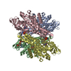
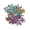
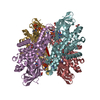
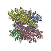
Crystal structure of isobutyryl-CoA dehydrogenase complexed with substrate/ligand.
Components
Acyl-CoA dehydrogenase family member 8, mitochondrial
Keywords
OXIDOREDUCTASE / flavoprotein / dehydrogenase / coenzyme A
Function / homology
Function and homology information
Oxidoreductases; Acting on the CH-CH group of donors; With a flavin as acceptor / valine catabolic process / acyl-CoA dehydrogenase activity / branched-chain amino acid catabolic process / Branched-chain amino acid catabolism / lipid metabolic process / flavin adenine dinucleotide binding / mitochondrial matrix Similarity search - Function
A: Acyl-CoA dehydrogenase family member 8, mitochondrial B: Acyl-CoA dehydrogenase family member 8, mitochondrial C: Acyl-CoA dehydrogenase family member 8, mitochondrial D: Acyl-CoA dehydrogenase family member 8, mitochondrial hetero molecules
In the structure databanks used in Yorodumi, some data are registered as the other names, "COVID-19 virus" and "2019-nCoV". Here are the details of the virus and the list of structure data.
Jan 31, 2019. EMDB accession codes are about to change! (news from PDBe EMDB page)
EMDB accession codes are about to change! (news from PDBe EMDB page)
The allocation of 4 digits for EMDB accession codes will soon come to an end. Whilst these codes will remain in use, new EMDB accession codes will include an additional digit and will expand incrementally as the available range of codes is exhausted. The current 4-digit format prefixed with “EMD-” (i.e. EMD-XXXX) will advance to a 5-digit format (i.e. EMD-XXXXX), and so on. It is currently estimated that the 4-digit codes will be depleted around Spring 2019, at which point the 5-digit format will come into force.
The EM Navigator/Yorodumi systems omit the EMD- prefix.
Related info.:Q: What is EMD? / ID/Accession-code notation in Yorodumi/EM Navigator
Yorodumi is a browser for structure data from EMDB, PDB, SASBDB, etc.
This page is also the successor to EM Navigator detail page, and also detail information page/front-end page for Omokage search.
The word "yorodu" (or yorozu) is an old Japanese word meaning "ten thousand". "mi" (miru) is to see.
Related info.:EMDB / PDB / SASBDB / Comparison of 3 databanks / Yorodumi Search / Aug 31, 2016. New EM Navigator & Yorodumi / Yorodumi Papers / Jmol/JSmol / Function and homology information / Changes in new EM Navigator and Yorodumi
 Movie
Movie Controller

 Yorodumi
 Open data
 Basic information
 Components
 Keywords
 OXIDOREDUCTASE /
  Function and homology information
 Authors
 Citation
 Structure visualization
 Downloads & links
 Other downloads
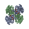

 PDBj
PDBj

 Assembly
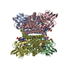


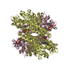
 Mass: 62.068 Da / Num. of mol.: 11 / Source method: obtained synthetically / Formula: C2H6O2
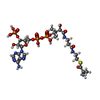
 Mass: 785.550 Da / Num. of mol.: 4 / Source method: obtained synthetically / Formula: C27H33N9O15P2 / Comment: FAD*YM
 Mass: 60.052 Da / Num. of mol.: 7 / Source method: obtained synthetically / Formula: C2H4O2
 Mass: 835.608 Da / Num. of mol.: 2 / Source method: obtained synthetically / Formula: C25H40N7O17P3S
 Sample preparation
 / Beamline: 14-BM-C / Wavelength: 0.9 Å
 Processing