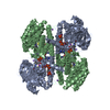
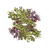
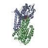
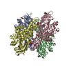
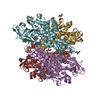
Entry Database : PDB / ID : 5lnxTitle Crystal structure of MmgC, an acyl-CoA dehydrogenase from bacillus subtilis. Acyl-CoA dehydrogenase Keywords / Function / homology Function Domain/homology Component
/ / / / / / / / / / / / / / / / / / / / / / / / / / / / / / / / / / Biological species Bacillus subtilis (bacteria)Method / / Resolution : 2.6 Å Authors Baker, G.E. / Race, P.R. Journal : To Be Published Title : Crystal structure of acyl-CoA dehydrogenase (MmgC) from bacillus subtilis.Authors : Baker, G.E. / Race, P.R. History Deposition Aug 7, 2016 Deposition site / Processing site Revision 1.0 Aug 16, 2017 Provider / Type Revision 1.1 Oct 16, 2019 Group / Category Revision 1.2 May 8, 2024 Group / Database references / Category / chem_comp_bond / database_2Item / _database_2.pdbx_database_accession
Show all Show less
 Movie
Movie Controller

 Yorodumi
 Open data
 Basic information
 Components
 Keywords
 Function and homology information
 Authors
 Citation
 Structure visualization
 Downloads & links
 Other downloads
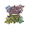


 PDBj
PDBj Assembly
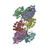

 Mass: 92.094 Da / Num. of mol.: 11 / Source method: obtained synthetically / Formula: C3H8O3
 Mass: 785.550 Da / Num. of mol.: 8 / Source method: obtained synthetically / Formula: C27H33N9O15P2 / Comment: FAD*YM
 Mass: 18.015 Da / Num. of mol.: 345 / Source method: isolated from a natural source / Formula: H2O
 Sample preparation
 / Beamline: I03 / Wavelength: 0.9763 Å
 Processing