Protocol: SINGLE WAVELENGTH / Monochromatic (M) / Laue (L): M / Scattering type: x-ray
Radiation wavelength
Wavelength: 1.541 Å / Relative weight: 1
Reflection
Redundancy: 5 % / Av σ(I) over netI: 17.5 / Number: 102082 / Rmerge(I) obs: 0.056 / Χ2: 1.26 / D res high: 1.65 Å / D res low: 50 Å / Num. obs: 20295 / % possible obs: 99.1
Diffraction reflection shell
Highest resolution (Å)
Lowest resolution (Å)
% possible obs (%)
ID
Rmerge(I) obs
Chi squared
Redundancy
3.55
50
99.7
1
0.034
0.78
5
2.82
3.55
99.9
1
0.038
0.837
5.2
2.46
2.82
99.9
1
0.062
1.184
5.2
2.24
2.46
99.8
1
0.081
1.265
5.2
2.08
2.24
99.8
1
0.106
1.194
5.1
1.96
2.08
99.4
1
0.149
1.344
5.1
1.86
1.96
98.9
1
0.23
1.486
5
1.78
1.86
98.8
1
0.312
1.51
4.9
1.71
1.78
98
1
0.444
1.568
4.9
1.65
1.71
96.9
1
0.578
1.558
4.7
Reflection
Resolution: 1.65→50 Å / Num. obs: 20360 / % possible obs: 99.1 % / Redundancy: 5 % / Rmerge(I) obs: 0.056 / Χ2: 1.305 / Net I/σ(I): 17.4
Reflection shell
Resolution (Å)
Redundancy (%)
Rmerge(I) obs
Num. unique all
Χ2
Diffraction-ID
% possible all
1.65-1.71
4.7
0.586
1981
1.592
1
96.9
1.71-1.78
4.9
0.452
1976
1.614
1
98
1.78-1.86
4.9
0.315
2015
1.597
1
98.8
1.86-1.96
5
0.234
1988
1.585
1
98.9
1.96-2.08
5.1
0.151
2021
1.351
1
99.4
2.08-2.24
5.1
0.106
2026
1.197
1
99.8
2.24-2.46
5.2
0.081
2056
1.284
1
99.8
2.46-2.82
5.2
0.062
2048
1.185
1
99.9
2.82-3.55
5.2
0.039
2074
0.881
1
99.9
3.55-50
5
0.035
2175
0.892
1
99.7
-
Phasing
Phasing
Method: MAD
Phasing MAD set site
ID
Cartn x (Å)
Cartn y (Å)
Cartn z (Å)
Atom type symbol
B iso
Occupancy
1
4.67
44.909
18.141
S
12.86
0.94
2
15.01
26.038
27.393
S
30
0.63
3
4.366
40.323
14.079
S
30.41
1.09
4
7.041
28.758
32.599
S
28.15
0.74
5
6.561
42.979
17.575
S
29.74
1.2
6
6.257
47.058
18.721
S
25.67
0.93
7
3.277
44.292
19.764
S
25.53
1.32
8
4.033
45.109
16.113
S
11.67
0.67
Phasing MR
Highest resolution
Lowest resolution
Rotation
1.91 Å
27.32 Å
Translation
1.91 Å
27.32 Å
-
Processing
Software
Name
Version
Classification
NB
DENZO
datareduction
SCALEPACK
datascaling
MOLREP
phasing
SHARP
phasing
SOLOMON
phasing
REFMAC
refinement
PDB_EXTRACT
2
dataextraction
CrystalClear
datacollection
HKL-2000
datareduction
Refinement
Method to determine structure: MOLECULAR REPLACEMENT / Resolution: 1.65→27.32 Å / Cor.coef. Fo:Fc: 0.966 / Cor.coef. Fo:Fc free: 0.959 / SU B: 1.176 / SU ML: 0.041 / Cross valid method: THROUGHOUT / σ(F): 0 / ESU R: 0.064 / ESU R Free: 0.065 / Stereochemistry target values: MAXIMUM LIKELIHOOD / Details: HYDROGENS HAVE BEEN ADDED IN THE RIDING POSITIONS
Rfactor
Num. reflection
% reflection
Selection details
Rfree
0.195
1044
5.1 %
RANDOM
Rwork
0.177
-
-
-
obs
0.177
20344
99.13 %
-
Solvent computation
Ion probe radii: 0.8 Å / Shrinkage radii: 0.8 Å / VDW probe radii: 1.4 Å / Solvent model: MASK
Displacement parameters
Biso mean: 24.562 Å2
Baniso -1
Baniso -2
Baniso -3
1-
0.89 Å2
0.44 Å2
0 Å2
2-
-
0.89 Å2
0 Å2
3-
-
-
-1.33 Å2
Refinement step
Cycle: LAST / Resolution: 1.65→27.32 Å
Protein
Nucleic acid
Ligand
Solvent
Total
Num. atoms
583
0
49
110
742
Refine LS restraints
Refine-ID
Type
Dev ideal
Dev ideal target
Number
X-RAY DIFFRACTION
r_bond_refined_d
0.02
0.021
651
X-RAY DIFFRACTION
r_angle_refined_deg
1.974
1.999
881
X-RAY DIFFRACTION
r_dihedral_angle_1_deg
5.117
5
70
X-RAY DIFFRACTION
r_dihedral_angle_2_deg
32.509
23.226
31
X-RAY DIFFRACTION
r_dihedral_angle_3_deg
14.078
15
112
X-RAY DIFFRACTION
r_dihedral_angle_4_deg
17.806
15
5
X-RAY DIFFRACTION
r_chiral_restr
0.145
0.2
88
X-RAY DIFFRACTION
r_gen_planes_refined
0.008
0.02
467
X-RAY DIFFRACTION
r_nbd_refined
0.2
0.2
285
X-RAY DIFFRACTION
r_nbtor_refined
0.31
0.2
419
X-RAY DIFFRACTION
r_xyhbond_nbd_refined
0.159
0.2
75
X-RAY DIFFRACTION
r_symmetry_vdw_refined
0.17
0.2
34
X-RAY DIFFRACTION
r_symmetry_hbond_refined
0.228
0.2
16
X-RAY DIFFRACTION
r_mcbond_it
1.236
1.5
357
X-RAY DIFFRACTION
r_mcangle_it
2.02
2
566
X-RAY DIFFRACTION
r_scbond_it
3.284
3
350
X-RAY DIFFRACTION
r_scangle_it
5.496
4.5
314
LS refinement shell
Resolution: 1.65→1.693 Å / Total num. of bins used: 20
Rfactor
Num. reflection
% reflection
Rfree
0.381
65
-
Rwork
0.351
1407
-
obs
-
1472
97.42 %
+
About Yorodumi
-
News
-
Feb 9, 2022. New format data for meta-information of EMDB entries
New format data for meta-information of EMDB entries
Version 3 of the EMDB header file is now the official format.
The previous official version 1.9 will be removed from the archive.
In the structure databanks used in Yorodumi, some data are registered as the other names, "COVID-19 virus" and "2019-nCoV". Here are the details of the virus and the list of structure data.
Jan 31, 2019. EMDB accession codes are about to change! (news from PDBe EMDB page)
EMDB accession codes are about to change! (news from PDBe EMDB page)
The allocation of 4 digits for EMDB accession codes will soon come to an end. Whilst these codes will remain in use, new EMDB accession codes will include an additional digit and will expand incrementally as the available range of codes is exhausted. The current 4-digit format prefixed with “EMD-” (i.e. EMD-XXXX) will advance to a 5-digit format (i.e. EMD-XXXXX), and so on. It is currently estimated that the 4-digit codes will be depleted around Spring 2019, at which point the 5-digit format will come into force.
The EM Navigator/Yorodumi systems omit the EMD- prefix.
Related info.:Q: What is EMD? / ID/Accession-code notation in Yorodumi/EM Navigator
Yorodumi is a browser for structure data from EMDB, PDB, SASBDB, etc.
This page is also the successor to EM Navigator detail page, and also detail information page/front-end page for Omokage search.
The word "yorodu" (or yorozu) is an old Japanese word meaning "ten thousand". "mi" (miru) is to see.
Related info.:EMDB / PDB / SASBDB / Comparison of 3 databanks / Yorodumi Search / Aug 31, 2016. New EM Navigator & Yorodumi / Yorodumi Papers / Jmol/JSmol / Function and homology information / Changes in new EM Navigator and Yorodumi
 Movie
Movie Controller

 Yorodumi
 Open data
 Basic information
 Components
 Keywords
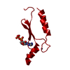
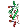
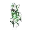
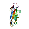
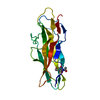
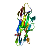
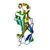
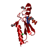
 DE NOVO PROTEIN / alpha/beta fold
 Function and homology information
 Authors
 Citation
 Structure visualization
 Downloads & links
 Other downloads
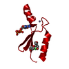


 PDBj
PDBj
 Assembly
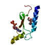


 Mass: 65.409 Da / Num. of mol.: 1 / Source method: obtained synthetically / Formula: Zn
 Mass: 35.453 Da / Num. of mol.: 1 / Source method: obtained synthetically / Formula: Cl
 Mass: 507.181 Da / Num. of mol.: 1 / Source method: obtained synthetically / Formula: C10H16N5O13P3 / Comment: ATP, energy-carrying molecule*YM
 Mass: 238.278 Da / Num. of mol.: 1 / Source method: obtained synthetically / Formula: C10H22O6 / Comment: precipitant*YM
 Sample preparation
 Processing