 Movie
Movie Controller

[English] 日本語
 Yorodumi
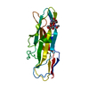
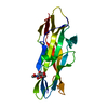
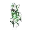
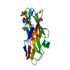
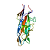
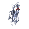
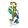
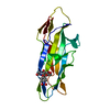
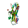
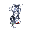
- PDB-2bs8: Crystal structure of F17b-G in complex with N-acetyl-D-glucosamine -
+ Open data

- Basic information
| Entry | Database: PDB / ID: 2bs8 | ||||||
|---|---|---|---|---|---|---|---|
| Title | Crystal structure of F17b-G in complex with N-acetyl-D-glucosamine | ||||||
 Components Components | ADHESIN | ||||||
 Keywords Keywords | SUGAR BINDING PROTEIN /  LECTIN / FIMBRIAE / PROTEIN-SUGAR COMPLEX / SUGAR-BINDING PROTEIN LECTIN / FIMBRIAE / PROTEIN-SUGAR COMPLEX / SUGAR-BINDING PROTEIN | ||||||
| Function / homology |  Function and homology information Function and homology informationadhesion of symbiont to host / pilus / carbohydrate binding / cell adhesionSimilarity search - Function | ||||||
| Biological species |  ESCHERICHIA COLI B (bacteria) ESCHERICHIA COLI B (bacteria) | ||||||
| Method | X-RAY DIFFRACTION / SYNCHROTRON / MOLECULAR REPLACEMENT / Resolution: 2.25 Å | ||||||
 Authors Authors | Buts, L. / Wellens, A. / VanMolle, I. / Wyns, L. / Loris, R. / Lahmann, M. / Oscarson, S. / DeGreve, H. / Bouckaert, J. | ||||||
 Citation Citation | Journal: Acta Crystallogr.,Sect.D / Year: 2005 Title: Impact of Natural Variation in Bacterial F17G Adhesins on Crystallization Behaviour. Authors: Buts, L. / Wellens, A. / Van Molle, I. / Wyns, L. / Loris, R. / Lahmann, M. / Oscarson, S. / De Greve, H. / Bouckaert, J. | ||||||
| History |
| ||||||
| Remark 700 | SHEET THE SHEET STRUCTURE OF THIS MOLECULE IS BIFURCATED. IN ORDER TO REPRESENT THIS FEATURE IN ... SHEET THE SHEET STRUCTURE OF THIS MOLECULE IS BIFURCATED. IN ORDER TO REPRESENT THIS FEATURE IN THE SHEET RECORDS BELOW, TWO SHEETS ARE DEFINED. |
- Structure visualization
| Structure viewer | Molecule: MolmilJmol/JSmol |
|---|
- Downloads & links
-Download
| PDBx/mmCIF format | 2bs8.cif.gz | 53.8 KB | Display | PDBx/mmCIF format |
|---|---|---|---|---|
| PDB format | pdb2bs8.ent.gz | 36.7 KB | Display | PDB format |
| PDBx/mmJSON format | 2bs8.json.gz | Tree view | PDBx/mmJSON format | |
| Others |  Other downloads Other downloads |
-Validation report
| Arichive directory | https://data.pdbj.org/pub/pdb/validation_reports/bs/2bs8ftp://data.pdbj.org/pub/pdb/validation_reports/bs/2bs8 | HTTPS FTP |
|---|
-Related structure data
| Related structure data |  1zk5C  1zplC  2bs7C  2bsbC 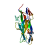 2bscC 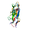 1o9wS C: citing same article ( S: Starting model for refinement |
|---|---|
| Similar structure data |
-Links
 PDBj
PDBj- Assembly
| Deposited unit | 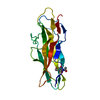
| ||||||||
|---|---|---|---|---|---|---|---|---|---|
| 1 |
| ||||||||
| Unit cell |
|
-Components
| #1: Protein | Mass: 18896.871 Da / Num. of mol.: 1 / Fragment: LECTIN DOMAIN, RESIDUES 23-198 Source method: isolated from a genetically manipulated source Source: (gene. exp.) ESCHERICHIA COLI B (bacteria) / Description: F17-POSITIVE E. COLI ISOLATE STRAIN / Plasmid: PBAD / Production host: ESCHERICHIA COLI (E. coli) / Strain (production host): BL21(DE3) / Variant (production host): C43 / References: UniProt: Q47200 |
|---|---|
| #2: Sugar | ChemComp-NAG / N-Acetylglucosamine  Type: D-saccharide, beta linking / Mass: 221.208 Da / Num. of mol.: 1 Type: D-saccharide, beta linking / Mass: 221.208 Da / Num. of mol.: 1Source method: isolated from a genetically manipulated source Formula: C8H15NO6 |
| #3: Water | ChemComp-HOH / Water Mass: 18.015 Da / Num. of mol.: 199 / Source method: isolated from a natural source / Formula: H2O Mass: 18.015 Da / Num. of mol.: 199 / Source method: isolated from a natural source / Formula: H2O |
| Sequence details | ELECTRON DENSITY AND NUCLEOTIDE |
-Experimental details
-Experiment
| Experiment | Method: X-RAY DIFFRACTION / Number of used crystals: 1 |
|---|
- Sample preparation
| Crystal | Density Matthews: 3 Å3/Da / Density % sol: 60 % |
|---|---|
| Crystal grow | Method: vapor diffusion, hanging drop Details: HANGING DROP: 1 MICROLITER OF 10% ETHANOL WITH 1.5 M NACL PLUS 1 MICROLITER OF 16 MG/ML F17BG |
-Data collection
| Diffraction | Mean temperature: 100 K |
|---|---|
| Diffraction source | Source: SYNCHROTRON / Site: EMBL/DESY, HAMBURG  / Beamline: X11 / Wavelength: 0.81 / Beamline: X11 / Wavelength: 0.81 |
| Detector | Type: MARRESEARCH / Detector: CCD / Date: Nov 20, 2002 / Details: BENT MIRROR |
| Radiation | Monochromator: TRIANGULAR MONOCHROMATOR / Protocol: SINGLE WAVELENGTH / Monochromatic (M) / Laue (L): M / Scattering type: x-ray |
| Radiation wavelength | Wavelength: 0.81 Å / Relative weight: 1 |
| Reflection | Resolution: 2.24→23 Å / Num. obs: 11000 / % possible obs: 96.8 % / Observed criterion σ(I): 0 / Redundancy: 5.1 % / Biso Wilson estimate: 43.9 Å2 / Rmerge(I) obs: 0.12 / Net I/σ(I): 10 |
| Reflection shell | Resolution: 2.24→2.32 Å / Redundancy: 4.5 % / Rmerge(I) obs: 0.21 / Mean I/σ(I) obs: 5 / % possible all: 91.1 |
- Processing
| Software |
| ||||||||||||||||||||||||||||||||||||||||||||||||||||||||||||
|---|---|---|---|---|---|---|---|---|---|---|---|---|---|---|---|---|---|---|---|---|---|---|---|---|---|---|---|---|---|---|---|---|---|---|---|---|---|---|---|---|---|---|---|---|---|---|---|---|---|---|---|---|---|---|---|---|---|---|---|---|---|
| Refinement | Method to determine structure: MOLECULAR REPLACEMENT Starting model: PDB ENTRY 1O9W Resolution: 2.25→22.91 Å / Rfactor Rfree error: 0.011 / Data cutoff high absF: 2439377.41 / Cross valid method: THROUGHOUT / σ(F): 0 / Stereochemistry target values: MAXIMUM LIKELIHOOD
| ||||||||||||||||||||||||||||||||||||||||||||||||||||||||||||
| Solvent computation | Solvent model: CNS BULK SOLVENT MODEL USED / Bsol: 216.269 Å2 / ksol: 0.466888 e/Å3 | ||||||||||||||||||||||||||||||||||||||||||||||||||||||||||||
| Displacement parameters | Biso mean: 35.35 Å2
| ||||||||||||||||||||||||||||||||||||||||||||||||||||||||||||
| Refine analyze |
| ||||||||||||||||||||||||||||||||||||||||||||||||||||||||||||
| Refinement step | Cycle: LAST / Resolution: 2.25→22.91 Å
| ||||||||||||||||||||||||||||||||||||||||||||||||||||||||||||
| Refine LS restraints |
| ||||||||||||||||||||||||||||||||||||||||||||||||||||||||||||
| LS refinement shell | Resolution: 2.25→2.32 Å / Total num. of bins used: 10 / % reflection obs: 91.1 % |
