 Movie
Movie Controller

[English] 日本語
 Yorodumi
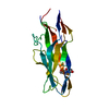
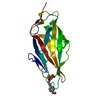
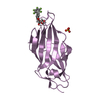
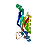
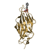
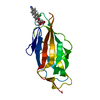
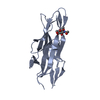
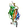
- PDB-1o9w: F17-aG lectin domain from Escherichia coli in complex with N-acet... -
+ Open data

- Basic information
| Entry | Database: PDB / ID: 1o9w | ||||||
|---|---|---|---|---|---|---|---|
| Title | F17-aG lectin domain from Escherichia coli in complex with N-acetyl-glucosamine | ||||||
 Components Components | F17A-G FIMBRIAL ADHESIN | ||||||
 Keywords Keywords | SUGAR BINDING PROTEIN /  BACTERIAL ADHESIN / BACTERIAL ATTACHMENT / SUGAR BINDING PROTEIN PATHOGENESIS / IMMUNOGLOBULIN FOLD BACTERIAL ADHESIN / BACTERIAL ATTACHMENT / SUGAR BINDING PROTEIN PATHOGENESIS / IMMUNOGLOBULIN FOLD | ||||||
| Function / homology |  Function and homology information Function and homology informationadhesion of symbiont to host / cell adhesion involved in single-species biofilm formation / pilus / carbohydrate bindingSimilarity search - Function | ||||||
| Biological species |  ESCHERICHIA COLI (E. coli) ESCHERICHIA COLI (E. coli) | ||||||
| Method | X-RAY DIFFRACTION / SYNCHROTRON / MOLECULAR REPLACEMENT / Resolution: 1.65 Å | ||||||
 Authors Authors | Buts, L. / De Genst, E. / Loris, R. / Oscarson, S. / Lahmann, M. / Messens, J. / Brosens, E. / Wyns, L. / Bouckaert, J. / De Greve, H. | ||||||
 Citation Citation | Journal: Mol.Microbiol. / Year: 2003 Title: The Fimbrial Adhesin F17-G of Enterotoxigenic Escherichia Coli Has an Immunoglobulin-Like Lectin Domain that Binds N-Acetylglucosamine Authors: Buts, L. / Bouckaert, J. / De Genst, E. / Loris, R. / Oscarson, S. / Lahmann, M. / Messens, J. / Brosens, E. / Wyns, L. / De Greve, H. #1: Journal: Acta Crystallogr.,Sect.D / Year: 2003 Title: Solving the Phase Problem for Carbohydrate -Binding Proteins Using Selenium Derivatives of Their Ligands: A Case Study Involving the Bacterial F17-G Adhesin Authors: Buts, L. / Loris, R. / De Genst, E. / Oscarson, S. / Lahmann, M. / Messens, J. / Brosens, E. / Wyns, L. / De Greve, H. / Bouckaert, J. #2: Journal: J.Bacteriol. / Year: 1991 Title: Identification, Characterization and Nucleotide Sequence of the F17G Gene, which Determines Receptor Binding of Escherichia Coli F17 Fimbriae Authors: Lintermans, P.F. / Bertels, A. / Schlicker, C. / Deboeck, F. / Charlier, G. / Pohl, P. / Norgren, M. / Normark, S. / Van Montagu, M. / De Greve, H. | ||||||
| History |
| ||||||
| Remark 700 | SHEET THE SHEET STRUCTURE OF THIS MOLECULE IS BIFURCATED. IN ORDER TO REPRESENT THIS FEATURE IN ... SHEET THE SHEET STRUCTURE OF THIS MOLECULE IS BIFURCATED. IN ORDER TO REPRESENT THIS FEATURE IN THE SHEET RECORDS BELOW, TWO SHEETS ARE DEFINED. |
- Structure visualization
| Structure viewer | Molecule: MolmilJmol/JSmol |
|---|
- Downloads & links
-Download
| PDBx/mmCIF format | 1o9w.cif.gz | 48.9 KB | Display | PDBx/mmCIF format |
|---|---|---|---|---|
| PDB format | pdb1o9w.ent.gz | 33.3 KB | Display | PDB format |
| PDBx/mmJSON format | 1o9w.json.gz | Tree view | PDBx/mmJSON format | |
| Others |  Other downloads Other downloads |
-Validation report
| Arichive directory | https://data.pdbj.org/pub/pdb/validation_reports/o9/1o9wftp://data.pdbj.org/pub/pdb/validation_reports/o9/1o9w | HTTPS FTP |
|---|
-Related structure data
| Related structure data |  1o9vSC 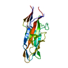 1o9zC S: Starting model for refinement C: citing same article ( |
|---|---|
| Similar structure data |


-Links
 PDBj
PDBj- Assembly
| Deposited unit | 
| ||||||||
|---|---|---|---|---|---|---|---|---|---|
| 1 |
| ||||||||
| Unit cell |
|
-Components
| #1: Protein | Mass: 19048.227 Da / Num. of mol.: 1 / Fragment: CARBOHYDRATE-BINDING DOMAIN, RESIDUES 23-199 Source method: isolated from a genetically manipulated source Source: (gene. exp.) ESCHERICHIA COLI (E. coli) / Plasmid: PHD52 / Production host: ESCHERICHIA COLI (E. coli) / Strain (production host): BL21 / References: UniProt: Q99003 |
|---|---|
| #2: Sugar | ChemComp-NAG / N-Acetylglucosamine  Type: D-saccharide, beta linking / Mass: 221.208 Da / Num. of mol.: 1 Type: D-saccharide, beta linking / Mass: 221.208 Da / Num. of mol.: 1Source method: isolated from a genetically manipulated source Formula: C8H15NO6 |
| #3: Water | ChemComp-HOH / Water Mass: 18.015 Da / Num. of mol.: 111 / Source method: isolated from a natural source / Formula: H2O Mass: 18.015 Da / Num. of mol.: 111 / Source method: isolated from a natural source / Formula: H2O |
-Experimental details
-Experiment
| Experiment | Method: X-RAY DIFFRACTION / Number of used crystals: 1 |
|---|
- Sample preparation
| Crystal | Density Matthews: 1.89 Å3/Da / Density % sol: 33 % | ||||||||||||||||||||||||||||||||||||||||||
|---|---|---|---|---|---|---|---|---|---|---|---|---|---|---|---|---|---|---|---|---|---|---|---|---|---|---|---|---|---|---|---|---|---|---|---|---|---|---|---|---|---|---|---|
| Crystal grow | pH: 4.6 Details: 30% PEG4000, 0.1M SODIUM ACETATE (PH 4.6), 0.2M AMMONIUM ACETATE | ||||||||||||||||||||||||||||||||||||||||||
| Crystal grow | *PLUS pH: 8 / Method: vapor diffusion | ||||||||||||||||||||||||||||||||||||||||||
| Components of the solutions | *PLUS
|
-Data collection
| Diffraction | Mean temperature: 100 K |
|---|---|
| Diffraction source | Source: SYNCHROTRON / Site: EMBL/DESY, HAMBURG  / Beamline: X13 / Wavelength: 0.8019 / Beamline: X13 / Wavelength: 0.8019 |
| Detector | Type: MARRESEARCH / Detector: CCD / Date: Aug 15, 2002 / Details: MIRRORS |
| Radiation | Monochromator: GRAPHITE / Protocol: SINGLE WAVELENGTH / Monochromatic (M) / Laue (L): M / Scattering type: x-ray |
| Radiation wavelength | Wavelength: 0.8019 Å / Relative weight: 1 |
| Reflection | Resolution: 1.65→25 Å / Num. obs: 20396 / % possible obs: 100 % / Redundancy: 48 % / Biso Wilson estimate: 27.8 Å2 / Rsym value: 0.073 / Net I/σ(I): 8.2 |
| Reflection shell | Resolution: 1.66→1.81 Å / Rsym value: 0.52 / % possible all: 99.9 |
- Processing
| Software |
| ||||||||||||||||||||||||||||||||||||||||||||||||||||||||||||
|---|---|---|---|---|---|---|---|---|---|---|---|---|---|---|---|---|---|---|---|---|---|---|---|---|---|---|---|---|---|---|---|---|---|---|---|---|---|---|---|---|---|---|---|---|---|---|---|---|---|---|---|---|---|---|---|---|---|---|---|---|---|
| Refinement | Method to determine structure: MOLECULAR REPLACEMENT Starting model: PDB ENTRY 1O9V Resolution: 1.65→24.77 Å / Rfactor Rfree error: 0.007 / Data cutoff high absF: 1557043.04 / Isotropic thermal model: RESTRAINED / Cross valid method: THROUGHOUT / σ(F): 0
| ||||||||||||||||||||||||||||||||||||||||||||||||||||||||||||
| Solvent computation | Solvent model: FLAT MODEL / Bsol: 61.5472 Å2 / ksol: 0.395503 e/Å3 | ||||||||||||||||||||||||||||||||||||||||||||||||||||||||||||
| Displacement parameters | Biso mean: 29.5 Å2
| ||||||||||||||||||||||||||||||||||||||||||||||||||||||||||||
| Refine analyze |
| ||||||||||||||||||||||||||||||||||||||||||||||||||||||||||||
| Refinement step | Cycle: LAST / Resolution: 1.65→24.77 Å
| ||||||||||||||||||||||||||||||||||||||||||||||||||||||||||||
| Refine LS restraints |
| ||||||||||||||||||||||||||||||||||||||||||||||||||||||||||||
| LS refinement shell | Resolution: 1.65→1.75 Å / Rfactor Rfree error: 0.024 / Total num. of bins used: 6
| ||||||||||||||||||||||||||||||||||||||||||||||||||||||||||||
| Xplor file |
| ||||||||||||||||||||||||||||||||||||||||||||||||||||||||||||
| Refine LS restraints | *PLUS
|
