 Movie
Movie Controller


+ Open data

- Basic information
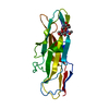
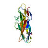
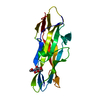
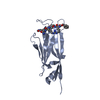
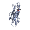
| Entry | Database: PDB / ID: 2cu9 | ||||||
|---|---|---|---|---|---|---|---|
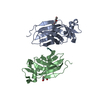
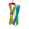
| Title | Crystal structure of Histone chaperone cia1 | ||||||
 Components Components | Histone chaperone cia1 | ||||||
 Keywords Keywords |  CHAPERONE / immunoglobulin fold / Structural Genomics / NPPSFA / National Project on Protein Structural and Functional Analyses / RIKEN Structural Genomics/Proteomics Initiative / RSGI CHAPERONE / immunoglobulin fold / Structural Genomics / NPPSFA / National Project on Protein Structural and Functional Analyses / RIKEN Structural Genomics/Proteomics Initiative / RSGI | ||||||
| Function / homology |  Function and homology information Function and homology informationH3-H4 histone complex chaperone activity / histone chaperone activity / DNA replication-dependent chromatin assembly / nucleosome disassembly / nucleosome assembly / chromatin organization / histone binding / chromatin remodeling / chromatin / nucleusSimilarity search - Function | ||||||
| Biological species |  Schizosaccharomyces pombe (fission yeast) Schizosaccharomyces pombe (fission yeast) | ||||||
| Method | X-RAY DIFFRACTION / SYNCHROTRON / MOLECULAR REPLACEMENT / Resolution: 1.8 Å | ||||||
 Authors Authors | Padmanabhan, B. / Yokoyama, S. / RIKEN Structural Genomics/Proteomics Initiative (RSGI) | ||||||
 Citation Citation | Journal: J.Biol.Chem. / Year: 2008 Title: Crystal Structures of Fission Yeast Histone Chaperone Asf1 Complexed with the Hip1 B-domain or the Cac2 C Terminus Authors: Malay, A.D. / Umehara, T. / Matsubara-Malay, K. / Padmanabhan, B. / Yokoyama, S. | ||||||
| History |
|
- Structure visualization
| Structure viewer | Molecule: MolmilJmol/JSmol |
|---|
- Downloads & links
-Download
| PDBx/mmCIF format | 2cu9.cif.gz | 51.8 KB | Display | PDBx/mmCIF format |
|---|---|---|---|---|
| PDB format | pdb2cu9.ent.gz | 35.7 KB | Display | PDB format |
| PDBx/mmJSON format | 2cu9.json.gz | Tree view | PDBx/mmJSON format | |
| Others |  Other downloads Other downloads |
-Validation report
| Arichive directory | https://data.pdbj.org/pub/pdb/validation_reports/cu/2cu9ftp://data.pdbj.org/pub/pdb/validation_reports/cu/2cu9 | HTTPS FTP |
|---|
-Related structure data
| Related structure data | 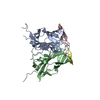 2z34C 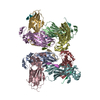 2z3fC 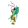 1wg3S C: citing same article ( S: Starting model for refinement |
|---|---|
| Similar structure data | |
| Other databases |

-Links
 PDBj
PDBj
- Assembly
| Deposited unit | 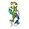
| ||||||||
|---|---|---|---|---|---|---|---|---|---|
| 1 |
| ||||||||
| Unit cell |
|
-Components
| #1: Protein | Mass: 18347.934 Da / Num. of mol.: 1 / Fragment: N-terminal region 1-161 Source method: isolated from a genetically manipulated source Source: (gene. exp.) Schizosaccharomyces pombe (fission yeast)Plasmid: pET15b / Production host:  Escherichia coli (E. coli) / References: UniProt: O74515 Escherichia coli (E. coli) / References: UniProt: O74515 |
|---|---|
| #2: Chemical | ChemComp-PG0 / 2-(2-Methoxyethoxy)ethanol  Mass: 120.147 Da / Num. of mol.: 1 / Source method: obtained synthetically / Formula: C5H12O3 / Comment: inhibitor, precipitant*YM Mass: 120.147 Da / Num. of mol.: 1 / Source method: obtained synthetically / Formula: C5H12O3 / Comment: inhibitor, precipitant*YM |
| #3: Water | ChemComp-HOH / Water Mass: 18.015 Da / Num. of mol.: 248 / Source method: isolated from a natural source / Formula: H2O Mass: 18.015 Da / Num. of mol.: 248 / Source method: isolated from a natural source / Formula: H2O |
-Experimental details
-Experiment
| Experiment | Method: X-RAY DIFFRACTION / Number of used crystals: 1 |
|---|
- Sample preparation
| Crystal | Density Matthews: 2.68 Å3/Da / Density % sol: 58 % |
|---|---|
| Crystal grow | Temperature: 293 K / Method: vapor diffusion / pH: 8 Details: PEG 6000, Ammonium Sulfate, pH 8.0, VAPOR DIFFUSION, temperature 293K |
-Data collection
| Diffraction | Mean temperature: 120 K |
|---|---|
| Diffraction source | Source: SYNCHROTRON / Site: SPring-8  / Beamline: BL26B1 / Wavelength: 1 Å / Beamline: BL26B1 / Wavelength: 1 Å |
| Detector | Type: RIGAKU JUPITER 210 / Detector: CCD / Date: Sep 24, 2004 |
| Radiation | Protocol: SINGLE WAVELENGTH / Monochromatic (M) / Laue (L): M / Scattering type: x-ray |
| Radiation wavelength | Wavelength: 1 Å / Relative weight: 1 |
| Reflection | Resolution: 1.8→50 Å / Num. obs: 17836 / % possible obs: 97.7 % / Observed criterion σ(I): -3 / Redundancy: 3.5 % / Rmerge(I) obs: 0.082 |
| Reflection shell | Resolution: 1.8→1.86 Å / Redundancy: 2.4 % / Rmerge(I) obs: 0.175 / Num. unique all: 1610 / % possible all: 87.4 |
- Processing
| Software |
| ||||||||||||||||||||||||||||||||||||||||||||||||||||||||||||||||||||||
|---|---|---|---|---|---|---|---|---|---|---|---|---|---|---|---|---|---|---|---|---|---|---|---|---|---|---|---|---|---|---|---|---|---|---|---|---|---|---|---|---|---|---|---|---|---|---|---|---|---|---|---|---|---|---|---|---|---|---|---|---|---|---|---|---|---|---|---|---|---|---|---|
| Refinement | Method to determine structure: MOLECULAR REPLACEMENT Starting model: PDB ENTRY 1WG3 Resolution: 1.8→20 Å / Cor.coef. Fo:Fc: 0.958 / Cor.coef. Fo:Fc free: 0.94 / SU B: 2.947 / SU ML: 0.092 / Cross valid method: THROUGHOUT / σ(F): 0 / ESU R: 0.133 / ESU R Free: 0.133 / Stereochemistry target values: MAXIMUM LIKELIHOOD
| ||||||||||||||||||||||||||||||||||||||||||||||||||||||||||||||||||||||
| Solvent computation | Ion probe radii: 0.8 Å / Shrinkage radii: 0.8 Å / VDW probe radii: 1.4 Å / Solvent model: BABINET MODEL WITH MASK | ||||||||||||||||||||||||||||||||||||||||||||||||||||||||||||||||||||||
| Displacement parameters | Biso mean: 33.102 Å2
| ||||||||||||||||||||||||||||||||||||||||||||||||||||||||||||||||||||||
| Refinement step | Cycle: LAST / Resolution: 1.8→20 Å
| ||||||||||||||||||||||||||||||||||||||||||||||||||||||||||||||||||||||
| Refine LS restraints |
| ||||||||||||||||||||||||||||||||||||||||||||||||||||||||||||||||||||||
| LS refinement shell | Resolution: 1.8→1.846 Å / Total num. of bins used: 20
|
