 Movie
Movie Controller


+ Open data

- Basic information
| Entry | Database: PDB / ID: 1wg3 | ||||||
|---|---|---|---|---|---|---|---|
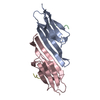
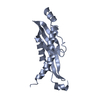
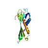
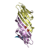
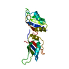
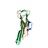
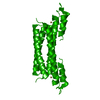
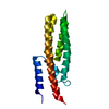
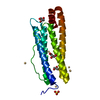
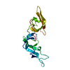
| Title | Structural analysis of yeast nucleosome-assembly factor CIA1p | ||||||
 Components Components | Anti-silencing protein 1 | ||||||
 Keywords Keywords |  REPLICATION / BETA-SANDWICH / RIKEN Structural Genomics/Proteomics Initiative / RSGI / Structural Genomics REPLICATION / BETA-SANDWICH / RIKEN Structural Genomics/Proteomics Initiative / RSGI / Structural Genomics | ||||||
| Function / homology |  Function and homology information: / Formation of Senescence-Associated Heterochromatin Foci (SAHF) / H3 histone acetyltransferase complex / acetyltransferase activator activity / DNA replication-dependent chromatin assembly / nucleosome disassembly / : / silent mating-type cassette heterochromatin formation / negative regulation of DNA damage checkpoint / subtelomeric heterochromatin formation ...: / Formation of Senescence-Associated Heterochromatin Foci (SAHF) / H3 histone acetyltransferase complex / acetyltransferase activator activity / DNA replication-dependent chromatin assembly / nucleosome disassembly / : / silent mating-type cassette heterochromatin formation / negative regulation of DNA damage checkpoint / subtelomeric heterochromatin formation / regulation of DNA repair / positive regulation of transcription elongation by RNA polymerase II / regulation of protein phosphorylation / nucleosome assembly / chromatin organization / histone binding / regulation of gene expression / chromosome, telomeric region / regulation of transcription by RNA polymerase II / nucleus / cytosol Function and homology information: / Formation of Senescence-Associated Heterochromatin Foci (SAHF) / H3 histone acetyltransferase complex / acetyltransferase activator activity / DNA replication-dependent chromatin assembly / nucleosome disassembly / : / silent mating-type cassette heterochromatin formation / negative regulation of DNA damage checkpoint / subtelomeric heterochromatin formation ...: / Formation of Senescence-Associated Heterochromatin Foci (SAHF) / H3 histone acetyltransferase complex / acetyltransferase activator activity / DNA replication-dependent chromatin assembly / nucleosome disassembly / : / silent mating-type cassette heterochromatin formation / negative regulation of DNA damage checkpoint / subtelomeric heterochromatin formation / regulation of DNA repair / positive regulation of transcription elongation by RNA polymerase II / regulation of protein phosphorylation / nucleosome assembly / chromatin organization / histone binding / regulation of gene expression / chromosome, telomeric region / regulation of transcription by RNA polymerase II / nucleus / cytosolSimilarity search - Function | ||||||
| Biological species |  Saccharomyces cerevisiae (brewer's yeast) Saccharomyces cerevisiae (brewer's yeast) | ||||||
| Method | X-RAY DIFFRACTION / SYNCHROTRON / MOLECULAR REPLACEMENT / Resolution: 3 Å | ||||||
 Authors Authors | Padmanabhan, B. / Kataoka, K. / Umehara, T. / Adachi, N. / Yokoyama, S. / Horikoshi, M. / RIKEN Structural Genomics/Proteomics Initiative (RSGI) | ||||||
 Citation Citation | Journal: J.Biochem.(Tokyo) / Year: 2005 Title: Structural Similarity between Histone Chaperone Cia1p/Asf1p and DNA-Binding Protein NF-{kappa}B Authors: Padmanabhan, B. / Kataoka, K. / Umehara, T. / Adachi, N. / Yokoyama, S. / Horikoshi, M. | ||||||
| History |
|
- Structure visualization
| Structure viewer | Molecule: MolmilJmol/JSmol |
|---|
- Downloads & links
-Download
| PDBx/mmCIF format | 1wg3.cif.gz | 48.7 KB | Display | PDBx/mmCIF format |
|---|---|---|---|---|
| PDB format | pdb1wg3.ent.gz | 33.4 KB | Display | PDB format |
| PDBx/mmJSON format | 1wg3.json.gz | Tree view | PDBx/mmJSON format | |
| Others |  Other downloads Other downloads |
-Validation report
| Arichive directory | https://data.pdbj.org/pub/pdb/validation_reports/wg/1wg3ftp://data.pdbj.org/pub/pdb/validation_reports/wg/1wg3 | HTTPS FTP |
|---|
-Related structure data
| Related structure data | 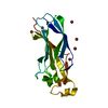 1rocS S: Starting model for refinement |
|---|---|
| Similar structure data | |
| Other databases |
-Links
 PDBj
PDBj- Assembly
| Deposited unit | 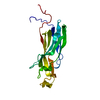
| ||||||||
|---|---|---|---|---|---|---|---|---|---|
| 1 |
| ||||||||
| Unit cell |
|
-Components
| #1: Protein | Mass: 19588.887 Da / Num. of mol.: 1 Source method: isolated from a genetically manipulated source Source: (gene. exp.) Saccharomyces cerevisiae (brewer's yeast)Plasmid: pGEX5X-2-CIA1-deltaC2 / Production host:  Escherichia coli (E. coli) / References: UniProt: P32447 Escherichia coli (E. coli) / References: UniProt: P32447 |
|---|---|
| #2: Water | ChemComp-HOH / Water Mass: 18.015 Da / Num. of mol.: 119 / Source method: isolated from a natural source / Formula: H2O Mass: 18.015 Da / Num. of mol.: 119 / Source method: isolated from a natural source / Formula: H2O |
-Experimental details
-Experiment
| Experiment | Method: X-RAY DIFFRACTION / Number of used crystals: 1 |
|---|
- Sample preparation
| Crystal | Density Matthews: 2.54 Å3/Da / Density % sol: 51 % |
|---|---|
| Crystal grow | Temperature: 293 K / Method: vapor diffusion, hanging drop / pH: 7.5 Details: PEG8K, AS, Tris-HCl, pH 7.5, VAPOR DIFFUSION, HANGING DROP, temperature 293K |
-Data collection
| Diffraction | Mean temperature: 298 K |
|---|---|
| Diffraction source | Source: SYNCHROTRON / Site: Photon Factory  / Beamline: BL-18B / Wavelength: 1 Å / Beamline: BL-18B / Wavelength: 1 Å |
| Detector | Type: ADSC QUANTUM 4 / Detector: CCD / Date: Oct 19, 2001 |
| Radiation | Protocol: SINGLE WAVELENGTH / Monochromatic (M) / Laue (L): M / Scattering type: x-ray |
| Radiation wavelength | Wavelength: 1 Å / Relative weight: 1 |
| Reflection | Resolution: 2.95→30 Å / Num. all: 4606 / Num. obs: 4606 / % possible obs: 99 % / Rmerge(I) obs: 0.085 / Net I/σ(I): 5.3 |
| Reflection shell | Resolution: 2.95→3.13 Å / Rmerge(I) obs: 0.287 / % possible all: 99 |
- Processing
| Software |
| |||||||||||||||||||||||||
|---|---|---|---|---|---|---|---|---|---|---|---|---|---|---|---|---|---|---|---|---|---|---|---|---|---|---|
| Refinement | Method to determine structure: MOLECULAR REPLACEMENT Starting model: 1ROC Resolution: 3→19.42 Å / Rfactor Rfree error: 0.019 / Data cutoff high absF: 7860443.03 / Data cutoff low absF: 0 / Isotropic thermal model: RESTRAINED / Cross valid method: THROUGHOUT / σ(F): 2 / Stereochemistry target values: Engh & Huber
| |||||||||||||||||||||||||
| Solvent computation | Solvent model: FLAT MODEL / Bsol: 70.9537 Å2 / ksol: 0.312976 e/Å3 | |||||||||||||||||||||||||
| Displacement parameters | Biso mean: 36.8 Å2
| |||||||||||||||||||||||||
| Refine analyze |
| |||||||||||||||||||||||||
| Refinement step | Cycle: LAST / Resolution: 3→19.42 Å
| |||||||||||||||||||||||||
| Refine LS restraints |
| |||||||||||||||||||||||||
| LS refinement shell | Resolution: 3→3.19 Å / Rfactor Rfree error: 0.062 / Total num. of bins used: 6
| |||||||||||||||||||||||||
| Xplor file |
|
