 Movie
Movie Controller

[English] 日本語
 Yorodumi
- PDB-5udo: Crystal structure of the coiled-coil domain from Listeria Innocua... -
+ Open data

- Basic information
| Entry | Database: PDB / ID: 5udo | ||||||
|---|---|---|---|---|---|---|---|
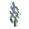
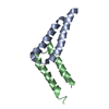
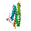
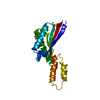
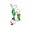
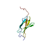
| Title | Crystal structure of the coiled-coil domain from Listeria Innocua Phage Integrase (Tetragonal Form II) | ||||||
 Components Components | A118 serine integrase | ||||||
 Keywords Keywords |  RECOMBINATION / site-specific recombination / coiled-coil RECOMBINATION / site-specific recombination / coiled-coil | ||||||
| Function / homology |  Function and homology information Function and homology informationDNA strand exchange activity / DNA integration / DNA binding / metal ion bindingSimilarity search - Function | ||||||
| Biological species |  Listeria innocua (bacteria) Listeria innocua (bacteria) | ||||||
| Method | X-RAY DIFFRACTION / SYNCHROTRON / MOLECULAR REPLACEMENT / Resolution: 2.541 Å | ||||||
 Authors Authors | Gupta, K. / Yuan, J.B. / Sharp, R. / Van Duyne, G.D. | ||||||
| Funding support |  United States, 1items United States, 1items
| ||||||
 Citation Citation | Journal: Nucleic Acids Res. / Year: 2017 Title: Coiled-coil interactions mediate serine integrase directionality. Authors: Gupta, K. / Sharp, R. / Yuan, J.B. / Li, H. / Van Duyne, G.D. #1: Journal: Nucleic Acids Res. / Year: 2013Title: Attachment site recognition and regulation of directionality by the serine integrases. Authors: Rutherford, K. / Yuan, P. / Perry, K. / Sharp, R. / Van Duyne, G.D. | ||||||
| History |
|
- Structure visualization
| Structure viewer | Molecule: MolmilJmol/JSmol |
|---|
- Downloads & links
-Download
| PDBx/mmCIF format | 5udo.cif.gz | 138.1 KB | Display | PDBx/mmCIF format |
|---|---|---|---|---|
| PDB format | pdb5udo.ent.gz | 91.6 KB | Display | PDB format |
| PDBx/mmJSON format | 5udo.json.gz | Tree view | PDBx/mmJSON format | |
| Others |  Other downloads Other downloads |
-Validation report
| Arichive directory | https://data.pdbj.org/pub/pdb/validation_reports/ud/5udoftp://data.pdbj.org/pub/pdb/validation_reports/ud/5udo | HTTPS FTP |
|---|
-Related structure data
| Related structure data |  5u96C  5uaeC  4kisS C: citing same article ( S: Starting model for refinement |
|---|---|
| Similar structure data |


-Links
 PDBj
PDBj- Assembly
| Deposited unit | 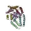
| ||||||||
|---|---|---|---|---|---|---|---|---|---|
| 1 | 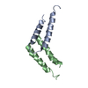
| ||||||||
| 2 | 
| ||||||||
| 3 | 
| ||||||||
| 4 | 
| ||||||||
| Unit cell |
|
-Components
| #1: Protein | Mass: 39155.633 Da / Num. of mol.: 8 / Fragment: coiled-coil domain (UNP residues 133-452) Source method: isolated from a genetically manipulated source Source: (gene. exp.) Listeria innocua (bacteria) / Gene: int / Production host: Escherichia coli BL21(DE3) (bacteria) / References: UniProt: Q928V6#2: Water | ChemComp-HOH / | Water Mass: 18.015 Da / Num. of mol.: 779 / Source method: isolated from a natural source / Formula: H2O Mass: 18.015 Da / Num. of mol.: 779 / Source method: isolated from a natural source / Formula: H2O |
|---|
-Experimental details
-Experiment
| Experiment | Method: X-RAY DIFFRACTION / Number of used crystals: 1 |
|---|
- Sample preparation
| Crystal grow | Temperature: 294.16 K / Method: vapor diffusion, hanging drop / pH: 7.5 / Details: 0.8-1.2 M Na Citrate |
|---|
-Data collection
| Diffraction | Mean temperature: 100 K |
|---|---|
| Diffraction source | Source: SYNCHROTRON / Site: CHESS / Beamline: F1 / Wavelength: 0.976 Å |
| Detector | Type: DECTRIS PILATUS3 2M / Detector: PIXEL / Date: Jun 3, 2016 |
| Radiation | Protocol: SINGLE WAVELENGTH / Monochromatic (M) / Laue (L): M / Scattering type: x-ray |
| Radiation wavelength | Wavelength: 0.976 Å / Relative weight: 1 |
| Reflection | Resolution: 2.541→50 Å / Num. obs: 16440 / % possible obs: 98.7 % / Redundancy: 5.5 % / Biso Wilson estimate: 67 Å2 / Rmerge(I) obs: 0.091 / Net I/σ(I): 12.9 |
| Reflection shell | Resolution: 2.541→2.64 Å / Rmerge(I) obs: 0.615 / Mean I/σ(I) obs: 1.95 / % possible all: 99.8 |
- Processing
| Software |
| |||||||||||||||||||||||||||||||||||||||||||||||||
|---|---|---|---|---|---|---|---|---|---|---|---|---|---|---|---|---|---|---|---|---|---|---|---|---|---|---|---|---|---|---|---|---|---|---|---|---|---|---|---|---|---|---|---|---|---|---|---|---|---|---|
| Refinement | Method to determine structure: MOLECULAR REPLACEMENT Starting model: 4KIS Resolution: 2.541→35.853 Å / Cross valid method: FREE R-VALUE / σ(F): 1.38 / Phase error: 32.79 / Stereochemistry target values: TWIN_LSQ_F
| |||||||||||||||||||||||||||||||||||||||||||||||||
| Solvent computation | Shrinkage radii: 0.9 Å / VDW probe radii: 1.11 Å / Solvent model: FLAT BULK SOLVENT MODEL | |||||||||||||||||||||||||||||||||||||||||||||||||
| Refinement step | Cycle: LAST / Resolution: 2.541→35.853 Å
| |||||||||||||||||||||||||||||||||||||||||||||||||
| Refine LS restraints |
| |||||||||||||||||||||||||||||||||||||||||||||||||
| LS refinement shell |
|
