 Movie
Movie Controller

[English] 日本語
 Yorodumi
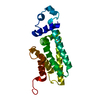
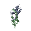
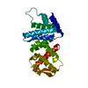
- PDB-1r1m: Structure of the OmpA-like domain of RmpM from Neisseria meningitidis -
+ Open data

- Basic information
| Entry | Database: PDB / ID: 1r1m | ||||||
|---|---|---|---|---|---|---|---|
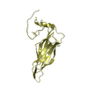
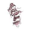
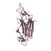
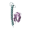
| Title | Structure of the OmpA-like domain of RmpM from Neisseria meningitidis | ||||||
 Components Components | Outer membrane protein class 4 | ||||||
 Keywords Keywords |  MEMBRANE PROTEIN MEMBRANE PROTEIN | ||||||
| Function / homology |  Function and homology informationporin activity / pore complex / monoatomic ion transport / cell outer membrane Function and homology informationporin activity / pore complex / monoatomic ion transport / cell outer membraneSimilarity search - Function | ||||||
| Biological species |  Neisseria meningitidis (bacteria) Neisseria meningitidis (bacteria) | ||||||
| Method | X-RAY DIFFRACTION / SAD / Resolution: 1.9 Å | ||||||
 Authors Authors | Grizot, S. / Buchanan, S.K. | ||||||
 Citation Citation | Journal: Mol.Microbiol. / Year: 2004 Title: Structure of the OmpA-like domain of RmpM from Neisseria meningitidis Authors: Grizot, S. / Buchanan, S.K. | ||||||
| History |
|
- Structure visualization
| Structure viewer | Molecule: MolmilJmol/JSmol |
|---|
- Downloads & links
-Download
| PDBx/mmCIF format | 1r1m.cif.gz | 38.6 KB | Display | PDBx/mmCIF format |
|---|---|---|---|---|
| PDB format | pdb1r1m.ent.gz | 29.5 KB | Display | PDB format |
| PDBx/mmJSON format | 1r1m.json.gz | Tree view | PDBx/mmJSON format | |
| Others |  Other downloads Other downloads |
-Validation report
| Arichive directory | https://data.pdbj.org/pub/pdb/validation_reports/r1/1r1mftp://data.pdbj.org/pub/pdb/validation_reports/r1/1r1m | HTTPS FTP |
|---|
-Related structure data
| Similar structure data |
|---|


-Links
 PDBj
PDBj- Assembly
| Deposited unit | 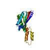
| ||||||||
|---|---|---|---|---|---|---|---|---|---|
| 1 |
| ||||||||
| Unit cell |
| ||||||||
| Components on special symmetry positions |
|
-Components
| #1: Protein | Mass: 18333.705 Da / Num. of mol.: 1 / Fragment: C-TERMINAL DOMAIN Source method: isolated from a genetically manipulated source Source: (gene. exp.) Neisseria meningitidis (bacteria) / Gene: RMPM / Plasmid: pET-20b / Production host: Escherichia coli (E. coli) / Strain (production host): Origami(DE3) / References: UniProt: P38367, UniProt: P0A0V3*PLUS |
|---|---|
| #2: Chemical | ChemComp-TRS / Tris  Mass: 122.143 Da / Num. of mol.: 1 / Source method: obtained synthetically / Formula: C4H12NO3 / Comment: pH buffer*YM Mass: 122.143 Da / Num. of mol.: 1 / Source method: obtained synthetically / Formula: C4H12NO3 / Comment: pH buffer*YM |
| #3: Water | ChemComp-HOH / Water Mass: 18.015 Da / Num. of mol.: 114 / Source method: isolated from a natural source / Formula: H2O Mass: 18.015 Da / Num. of mol.: 114 / Source method: isolated from a natural source / Formula: H2O |
-Experimental details
-Experiment
| Experiment | Method: X-RAY DIFFRACTION / Number of used crystals: 1 |
|---|
- Sample preparation
| Crystal | Density Matthews: 2.3 Å3/Da / Density % sol: 46.64 % |
|---|---|
| Crystal grow | Temperature: 294 K / Method: vapor diffusion, hanging drop / pH: 8 Details: PEG 1000, Tris, pH 8.0, VAPOR DIFFUSION, HANGING DROP, temperature 294K |
-Data collection
| Diffraction | Mean temperature: 95 K |
|---|---|
| Diffraction source | Source: ROTATING ANODE / Type: RIGAKU RU200 / Wavelength: 1.5418 Å |
| Detector | Type: RIGAKU RAXIS IV / Detector: IMAGE PLATE / Date: Nov 12, 2002 / Details: mirrors |
| Radiation | Protocol: SINGLE WAVELENGTH / Monochromatic (M) / Laue (L): M / Scattering type: x-ray |
| Radiation wavelength | Wavelength: 1.5418 Å / Relative weight: 1 |
| Reflection | Resolution: 1.9→50 Å / Num. all: 13605 / Num. obs: 13591 / % possible obs: 96.2 % / Observed criterion σ(F): 0 / Observed criterion σ(I): 0 / Redundancy: 4.8 % / Biso Wilson estimate: 20.2 Å2 / Rmerge(I) obs: 0.053 / Rsym value: 0.053 / Net I/σ(I): 25 |
| Reflection shell | Resolution: 1.9→1.97 Å / Redundancy: 4.8 % / Rmerge(I) obs: 0.254 / Mean I/σ(I) obs: 6 / Num. unique all: 1361 / Rsym value: 0.254 / % possible all: 98.8 |
- Processing
| Software |
| |||||||||||||||||||||||||
|---|---|---|---|---|---|---|---|---|---|---|---|---|---|---|---|---|---|---|---|---|---|---|---|---|---|---|
| Refinement | Method to determine structure: SAD / Resolution: 1.9→20 Å / Isotropic thermal model: isotropic / Cross valid method: THROUGHOUT / σ(F): 0 / Stereochemistry target values: Engh & Huber
| |||||||||||||||||||||||||
| Displacement parameters | Biso mean: 25.3 Å2 | |||||||||||||||||||||||||
| Refine analyze |
| |||||||||||||||||||||||||
| Refinement step | Cycle: LAST / Resolution: 1.9→20 Å
| |||||||||||||||||||||||||
| Refine LS restraints |
| |||||||||||||||||||||||||
| LS refinement shell | Resolution: 1.9→2.02 Å / Rfactor Rfree error: 0.024
|
