CELL INVASION / IPAD / T3SS / SEMET / INVASIN / VIRULENCE / SHIGELLA FLEXNERI / TYPE III SECRETION
Function / homology
Function and homology information
effector-mediated activation of programmed cell death in host / extracellular region Similarity search - Function
IpaD-like / IpaD-like / Type III secretion systems tip complex components / BipD-like superfamily / Type III secretion systems tip complex components / Up-down Bundle / Mainly Alpha Similarity search - Domain/homology
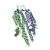
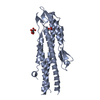
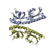
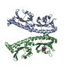
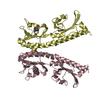
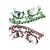
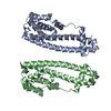
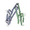
Journal: J Biol Chem / Year: 2007 Title: Self-chaperoning of the type III secretion system needle tip proteins IpaD and BipD. Authors: Steven Johnson / Pietro Roversi / Marianela Espina / Andrew Olive / Janet E Deane / Susan Birket / Terry Field / William D Picking / Ariel J Blocker / Edouard E Galyov / Wendy L Picking / Susan M Lea / Abstract: Bacteria expressing type III secretion systems (T3SS) have been responsible for the deaths of millions worldwide, acting as key virulence elements in diseases ranging from plague to typhoid fever. ...Bacteria expressing type III secretion systems (T3SS) have been responsible for the deaths of millions worldwide, acting as key virulence elements in diseases ranging from plague to typhoid fever. The T3SS is composed of a basal body, which traverses both bacterial membranes, and an external needle through which effector proteins are secreted. We report multiple crystal structures of two proteins that sit at the tip of the needle and are essential for virulence: IpaD from Shigella flexneri and BipD from Burkholderia pseudomallei. The structures reveal that the N-terminal domains of the molecules are intramolecular chaperones that prevent premature oligomerization, as well as sharing structural homology with proteins involved in eukaryotic actin rearrangement. Crystal packing has allowed us to construct a model for the tip complex that is supported by mutations designed using the structure.
History
Deposition
Nov 24, 2006
Deposition site: PDBE / Processing site: PDBE
Revision 1.0
Nov 30, 2006
Provider: repository / Type: Initial release
Revision 1.1
Apr 1, 2015
Group: Data collection / Database references ...Data collection / Database references / Derived calculations / Non-polymer description / Other / Source and taxonomy / Structure summary
Protocol: SINGLE WAVELENGTH / Monochromatic (M) / Laue (L): M / Scattering type: x-ray
Radiation wavelength
Wavelength: 0.9792 Å / Relative weight: 1
Reflection
Resolution: 3.1→33.9 Å / Num. obs: 62274 / % possible obs: 99.4 % / Observed criterion σ(I): 0 / Redundancy: 5.8 % / Biso Wilson estimate: 15.4 Å2 / Rmerge(I) obs: 0.1 / Net I/σ(I): 4.9
Reflection shell
Resolution: 3.1→3.27 Å / Redundancy: 6 % / Rmerge(I) obs: 0.31 / Mean I/σ(I) obs: 2.3 / % possible all: 99.4
-
Processing
Software
Name
Version
Classification
SOLOMON
modelbuilding
TNT
5.6.1
refinement
SCALA
datascaling
SHELXCD
phasing
SHELXD
phasing
SHARP
phasing
SOLOMON
phasing
RESOLVE
phasing
ARP/wARP
phasing
HELIXBUILD
phasing
Refinement
Method to determine structure: OTHER / Resolution: 3.1→34 Å / Isotropic thermal model: TNT BCORREL / Cross valid method: THROUGHOUT / σ(F): 0 / Stereochemistry target values: TNT PROTEGEO Details: REFINED IN BUSTER-TNT BETA 1.9.3 WAS USED AS A MOLECULAR REPLACEMENT MODEL FOR DETERMINING THE STRUCTURES OF PDB ENTRIES 2J0N, 2J0O
In the structure databanks used in Yorodumi, some data are registered as the other names, "COVID-19 virus" and "2019-nCoV". Here are the details of the virus and the list of structure data.
Jan 31, 2019. EMDB accession codes are about to change! (news from PDBe EMDB page)
EMDB accession codes are about to change! (news from PDBe EMDB page)
The allocation of 4 digits for EMDB accession codes will soon come to an end. Whilst these codes will remain in use, new EMDB accession codes will include an additional digit and will expand incrementally as the available range of codes is exhausted. The current 4-digit format prefixed with “EMD-” (i.e. EMD-XXXX) will advance to a 5-digit format (i.e. EMD-XXXXX), and so on. It is currently estimated that the 4-digit codes will be depleted around Spring 2019, at which point the 5-digit format will come into force.
The EM Navigator/Yorodumi systems omit the EMD- prefix.
Related info.:Q: What is EMD? / ID/Accession-code notation in Yorodumi/EM Navigator
Yorodumi is a browser for structure data from EMDB, PDB, SASBDB, etc.
This page is also the successor to EM Navigator detail page, and also detail information page/front-end page for Omokage search.
The word "yorodu" (or yorozu) is an old Japanese word meaning "ten thousand". "mi" (miru) is to see.
Related info.:EMDB / PDB / SASBDB / Comparison of 3 databanks / Yorodumi Search / Aug 31, 2016. New EM Navigator & Yorodumi / Yorodumi Papers / Jmol/JSmol / Function and homology information / Changes in new EM Navigator and Yorodumi
 Movie
Movie Controller


 Open data
 Basic information
 Components
 Keywords
 T3SS /
  Function and homology information
 Authors
 Citation
 Structure visualization
 Downloads & links
 Other downloads


 PDBj
PDBj Assembly
 Mass: 18.015 Da / Num. of mol.: 8 / Source method: isolated from a natural source / Formula: H2O
 Sample preparation
 / Beamline: ID14-1 / Wavelength: 0.9792
  Processing