 Movie
Movie Controller

[English] 日本語
 Yorodumi
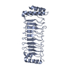
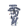
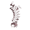
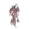
- PDB-1k24: Crystal Structure of the OpcA Outer Membrane Adhesin/Invasin from... -
+ Open data

- Basic information
| Entry | Database: PDB / ID: 1k24 | ||||||
|---|---|---|---|---|---|---|---|
| Title | Crystal Structure of the OpcA Outer Membrane Adhesin/Invasin from Neisseria meningitidis | ||||||
 Components Components | outer membrane protein Virulence-related outer membrane protein family Virulence-related outer membrane protein family | ||||||
 Keywords Keywords | MEMBRANE PROTEIN / Adhesin / Invasin / Outer Membrane / Beta Barrel | ||||||
| Function / homology |  Function and homology information Function and homology information | ||||||
| Biological species |  Neisseria meningitidis (bacteria) Neisseria meningitidis (bacteria) | ||||||
| Method | X-RAY DIFFRACTION / SYNCHROTRON / MIR / Resolution: 2.03 Å | ||||||
 Authors Authors | Prince, S.M. / Achtman, M. / Derrick, J.P. | ||||||
 Citation Citation | Journal: Proc.Natl.Acad.Sci.USA / Year: 2002 Title: Crystal structure of the OpcA integral membrane adhesin from Neisseria meningitidis. Authors: Prince, S.M. / Achtman, M. / Derrick, J.P. #1: Journal: Acta Crystallogr.,Sect.D / Year: 2001Title: Expression, Refolding and Crystallization of the OpcA Invasin from Neisseria meningitidis Authors: Prince, S.M. / Feron, C. / Janssens, D. / Lobet, Y. / Achtman, M. / Kusecek, B. / Bullough, P.A. / Derrick, J.P. #2: Journal: J.EXP.MED. / Year: 1988Title: Purification and Characterization of Eight Class 5 outer Membrane Protein Variants from a Clone of Neisseria meningitidis Serogroup A Authors: Achtman, M. / Neilbert, M. / Crowe, B.A. / Strittmatter, W. / Kusecek, B. / Weyse, E. / Walsh, M.J. / Slawig, B. / Morelli, G. / Moll, A. / Blake, M. #3: Journal: MOL.MICROBIOL. / Year: 1999Title: Projection Structure of Reconstituted Opc Outer Membrane Protein from Neisseria meningitidis Authors: Collins, R. / Achtman, M. / Ford, R. / Bullough, P. / Derrick, J. #4: Journal: MOL.MICROBIOL. / Year: 1997Title: Two-dimensional Structure of the Opc Invasin from Neisseria meningitidis Authors: Merker, P. / Tommassen, J. / Kusacek, B. / Virji, M. / Sesardic, D. / Achtman, M. | ||||||
| History |
|
- Structure visualization
| Structure viewer | Molecule: MolmilJmol/JSmol |
|---|
- Downloads & links
-Download
| PDBx/mmCIF format | 1k24.cif.gz | 68.1 KB | Display | PDBx/mmCIF format |
|---|---|---|---|---|
| PDB format | pdb1k24.ent.gz | 50 KB | Display | PDB format |
| PDBx/mmJSON format | 1k24.json.gz | Tree view | PDBx/mmJSON format | |
| Others |  Other downloads Other downloads |
-Validation report
| Arichive directory | https://data.pdbj.org/pub/pdb/validation_reports/k2/1k24ftp://data.pdbj.org/pub/pdb/validation_reports/k2/1k24 | HTTPS FTP |
|---|
-Related structure data
| Similar structure data |
|---|


-Links
 PDBj
PDBj- Assembly
| Deposited unit | 
| ||||||||
|---|---|---|---|---|---|---|---|---|---|
| 1 |
| ||||||||
| 2 | 
| ||||||||
| Unit cell |
| ||||||||
| Components on special symmetry positions |
|
-Components
| #1: Protein | Virulence-related outer membrane protein family / OpcA protein Mass: 28092.643 Da / Num. of mol.: 1 / Source method: isolated from a natural source / Details: Outer membrane / Source: (natural) Neisseria meningitidis (bacteria) / References: UniProt: Q9AE79, UniProt: Q51227*PLUS | ||||
|---|---|---|---|---|---|
| #2: Chemical |   Mass: 65.409 Da / Num. of mol.: 3 / Source method: obtained synthetically / Formula: Zn Mass: 65.409 Da / Num. of mol.: 3 / Source method: obtained synthetically / Formula: Zn#3: Chemical | Polyethylene glycol  Mass: 238.278 Da / Num. of mol.: 2 / Source method: obtained synthetically / Formula: C10H22O6 / Comment: precipitant*YM Mass: 238.278 Da / Num. of mol.: 2 / Source method: obtained synthetically / Formula: C10H22O6 / Comment: precipitant*YM#4: Water | ChemComp-HOH / | Water Mass: 18.015 Da / Num. of mol.: 144 / Source method: isolated from a natural source / Formula: H2O Mass: 18.015 Da / Num. of mol.: 144 / Source method: isolated from a natural source / Formula: H2O |
-Experimental details
-Experiment
| Experiment | Method: X-RAY DIFFRACTION / Number of used crystals: 1 |
|---|
- Sample preparation
| Crystal | Density Matthews: 3.04 Å3/Da / Density % sol: 59.2 % | ||||||||||||||||||||||||||||||||||||||||||||||||||||||||||||||||||||||
|---|---|---|---|---|---|---|---|---|---|---|---|---|---|---|---|---|---|---|---|---|---|---|---|---|---|---|---|---|---|---|---|---|---|---|---|---|---|---|---|---|---|---|---|---|---|---|---|---|---|---|---|---|---|---|---|---|---|---|---|---|---|---|---|---|---|---|---|---|---|---|---|
| Crystal grow | Temperature: 293 K / Method: vapor diffusion, hanging drop / pH: 7.5 Details: PEG 6K, Zinc Acetate, Zinc Chloride, C10E5, Beta-heptyl-glucoside, TRIS/HCl, pH 7.5, VAPOR DIFFUSION, HANGING DROP, temperature 293K | ||||||||||||||||||||||||||||||||||||||||||||||||||||||||||||||||||||||
| Crystal grow | *PLUS Details: Prince, S.M., (2001) Acta Crystallogr., Sect.D, 57, 1164. | ||||||||||||||||||||||||||||||||||||||||||||||||||||||||||||||||||||||
| Components of the solutions | *PLUS
|
-Data collection
| Diffraction | Mean temperature: 100 K |
|---|---|
| Diffraction source | Source: SYNCHROTRON / Site: ESRF  / Beamline: ID14-2 / Wavelength: 0.934 Å / Beamline: ID14-2 / Wavelength: 0.934 Å |
| Detector | Type: MARRESEARCH / Detector: CCD / Date: Oct 9, 1999 / Details: toroidal mirror |
| Radiation | Monochromator: Diamond & Ge-crystal / Protocol: SINGLE WAVELENGTH / Monochromatic (M) / Laue (L): M / Scattering type: x-ray |
| Radiation wavelength | Wavelength: 0.934 Å / Relative weight: 1 |
| Reflection | Resolution: 2.03→36.5 Å / Num. all: 21875 / Num. obs: 21875 / % possible obs: 98.3 % / Observed criterion σ(F): 0 / Observed criterion σ(I): -5 / Redundancy: 10.2 % / Biso Wilson estimate: 37.6 Å2 / Rmerge(I) obs: 0.084 / Rsym value: 0.084 / Net I/σ(I): 16.5 |
| Reflection shell | Resolution: 2.03→2.08 Å / Redundancy: 6.2 % / Rmerge(I) obs: 0.325 / Mean I/σ(I) obs: 4.1 / Num. unique all: 1320 / Rsym value: 0.325 / % possible all: 82.4 |
| Reflection | *PLUS Highest resolution: 2 Å / Lowest resolution: 36.4 Å / Rmerge(I) obs: 0.081 |
| Reflection shell | *PLUS Rmerge(I) obs: 0.325 |
- Processing
| Software |
| ||||||||||||||||||||||||||||||||||||||||||||||||||||
|---|---|---|---|---|---|---|---|---|---|---|---|---|---|---|---|---|---|---|---|---|---|---|---|---|---|---|---|---|---|---|---|---|---|---|---|---|---|---|---|---|---|---|---|---|---|---|---|---|---|---|---|---|---|
| Refinement | Method to determine structure: MIR / Resolution: 2.03→36.5 Å / SU B: 4.423 / SU ML: 0.125 Isotropic thermal model: Overall Anisotropic B-factor applied. 28 TLS tensors refined, Residual B-factors also refined. Cross valid method: THROUGHOUT / σ(F): 0 / ESU R: 0.203 / ESU R Free: 0.182 / Stereochemistry target values: Engh & Huber Details: Maximum Likelihood target, Bulk Solvent Correction applied. TLS TENSORS WERE USED TO MODEL DISORDER, THE DEPOSITION CONTAINS THE EQUIVALENT B-FACTORS CALCULATED FROM THESE TENSORS.
| ||||||||||||||||||||||||||||||||||||||||||||||||||||
| Displacement parameters | Biso mean: 70.2 Å2
| ||||||||||||||||||||||||||||||||||||||||||||||||||||
| Refine analyze |
| ||||||||||||||||||||||||||||||||||||||||||||||||||||
| Refinement step | Cycle: LAST / Resolution: 2.03→36.5 Å
| ||||||||||||||||||||||||||||||||||||||||||||||||||||
| Refine LS restraints |
| ||||||||||||||||||||||||||||||||||||||||||||||||||||
| LS refinement shell | Resolution: 2.03→2.129 Å
| ||||||||||||||||||||||||||||||||||||||||||||||||||||
| Refinement | *PLUS Rfactor all: 0.237 / Rfactor obs: 0.235 / Rfactor Rfree: 0.274 / Rfactor Rwork: 0.235 | ||||||||||||||||||||||||||||||||||||||||||||||||||||
| Solvent computation | *PLUS | ||||||||||||||||||||||||||||||||||||||||||||||||||||
| Displacement parameters | *PLUS | ||||||||||||||||||||||||||||||||||||||||||||||||||||
| Refine LS restraints | *PLUS
| ||||||||||||||||||||||||||||||||||||||||||||||||||||
| LS refinement shell | *PLUS Rfactor Rfree: 0.288 / Rfactor Rwork: 0.227 / Rfactor obs: 0.227 |
