 Movie
Movie Controller

[English] 日本語
 Yorodumi
- PDB-2i7n: Crystal structure of human PANK1 alpha: the catalytic core domain... -
+ Open data

- Basic information
| Entry | Database: PDB / ID: 2i7n | ||||||
|---|---|---|---|---|---|---|---|
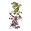
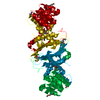
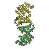
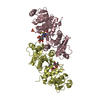
| Title | Crystal structure of human PANK1 alpha: the catalytic core domain in complex with AcCoA | ||||||
 Components Components | Pantothenate kinase 1 | ||||||
 Keywords Keywords | TRANSFERASE / PANK | ||||||
| Function / homology |  Function and homology information Function and homology informationCoenzyme A biosynthesis / acetyl-CoA binding / pantothenate kinase / pantothenate kinase activity / coenzyme A biosynthetic process / clathrin-coated vesicle / recycling endosome / phosphorylation / nucleolus / protein homodimerization activity ...Coenzyme A biosynthesis / acetyl-CoA binding / pantothenate kinase / pantothenate kinase activity / coenzyme A biosynthetic process / clathrin-coated vesicle / recycling endosome / phosphorylation / nucleolus / protein homodimerization activity / ATP binding / nucleus / cytosol / cytoplasmSimilarity search - Function | ||||||
| Biological species |  Homo sapiens (human) Homo sapiens (human) | ||||||
| Method | X-RAY DIFFRACTION / SYNCHROTRON / MAD / Resolution: 1.9 Å | ||||||
 Authors Authors | Hong, B.S. / Wang, L. / Tempel, W. / Loppnau, P. / Allali-Hassani, A. / Arrowsmith, C.H. / Edwards, A.M. / Sundstrom, M. / Weigelt, J. / Bochkarev, A. / Park, H.W. | ||||||
 Citation Citation | Journal: J.Biol.Chem. / Year: 2007 Title: Crystal structures of human pantothenate kinases. Insights into allosteric regulation and mutations linked to a neurodegeneration disorder. Authors: Hong, B.S. / Senisterra, G. / Rabeh, W.M. / Vedadi, M. / Leonardi, R. / Zhang, Y.M. / Rock, C.O. / Jackowski, S. / Park, H.W. | ||||||
| History |
|
- Structure visualization
| Structure viewer | Molecule: MolmilJmol/JSmol |
|---|
- Downloads & links
-Download
| PDBx/mmCIF format | 2i7n.cif.gz | 147.5 KB | Display | PDBx/mmCIF format |
|---|---|---|---|---|
| PDB format | pdb2i7n.ent.gz | 114.6 KB | Display | PDB format |
| PDBx/mmJSON format | 2i7n.json.gz | Tree view | PDBx/mmJSON format | |
| Others |  Other downloads Other downloads |
-Validation report
| Arichive directory | https://data.pdbj.org/pub/pdb/validation_reports/i7/2i7nftp://data.pdbj.org/pub/pdb/validation_reports/i7/2i7n | HTTPS FTP |
|---|
-Related structure data


-Links
 PDBj
PDBj- Assembly
| Deposited unit | 
| |||||||||
|---|---|---|---|---|---|---|---|---|---|---|
| 1 |
| |||||||||
| Unit cell |
| |||||||||
| Components on special symmetry positions |
| |||||||||
| Details | The biological assembly is a dimer in the asymmetric unit |
-Components
| #1: Protein | / Pantothenic acid kinase 1 / hPanK1 / hPanK Mass: 40176.188 Da / Num. of mol.: 2 / Fragment: residues 234-593 Source method: isolated from a genetically manipulated source Source: (gene. exp.) Homo sapiens (human) / Gene: PANK1, PANK / Plasmid: pET28aLIC / Species (production host): Escherichia coli / Production host:  Escherichia coli BL21(DE3) (bacteria) / Strain (production host): BL21(DE3) / References: UniProt: Q8TE04, pantothenate kinase Escherichia coli BL21(DE3) (bacteria) / Strain (production host): BL21(DE3) / References: UniProt: Q8TE04, pantothenate kinase#2: Chemical | Acetyl-CoA  Mass: 809.571 Da / Num. of mol.: 2 / Source method: obtained synthetically / Formula: C23H38N7O17P3S Mass: 809.571 Da / Num. of mol.: 2 / Source method: obtained synthetically / Formula: C23H38N7O17P3S#3: Water | ChemComp-HOH / | Water Mass: 18.015 Da / Num. of mol.: 252 / Source method: isolated from a natural source / Formula: H2O Mass: 18.015 Da / Num. of mol.: 252 / Source method: isolated from a natural source / Formula: H2O |
|---|
-Experimental details
-Experiment
| Experiment | Method: X-RAY DIFFRACTION / Number of used crystals: 2 |
|---|
- Sample preparation
| Crystal | Density Matthews: 3.07 Å3/Da / Density % sol: 59.95 % |
|---|---|
| Crystal grow | Temperature: 298 K / Method: vapor diffusion, sitting drop / pH: 7.5 Details: 25% PEG400, 0.2M CaCl, 0.1M Na HEPES, pH 7.5, VAPOR DIFFUSION, SITTING DROP, temperature 298K |
-Data collection
| Diffraction |
| ||||||||||||||||||
|---|---|---|---|---|---|---|---|---|---|---|---|---|---|---|---|---|---|---|---|
| Diffraction source | Source: SYNCHROTRON / Site: APS  / Beamline: 19-BM / Wavelength: 0.9 Å / Beamline: 19-BM / Wavelength: 0.9 Å | ||||||||||||||||||
| Detector | Type: SBC-3 / Detector: CCD / Date: Dec 19, 2005 | ||||||||||||||||||
| Radiation |
| ||||||||||||||||||
| Radiation wavelength | Wavelength: 0.9 Å / Relative weight: 1 | ||||||||||||||||||
| Reflection | Resolution: 1.9→50 Å / Num. obs: 75175 / % possible obs: 99.5 % / Observed criterion σ(F): 1 / Observed criterion σ(I): 1 / Redundancy: 7.6 % / Biso Wilson estimate: 17 Å2 / Rmerge(I) obs: 0.07 / Rsym value: 0.067 / Net I/σ(I): 47.6 | ||||||||||||||||||
| Reflection shell | Resolution: 1.9→1.97 Å / Redundancy: 5.2 % / Rmerge(I) obs: 0.38 / Mean I/σ(I) obs: 3.5 / Num. unique all: 7453 / Rsym value: 0.61 / % possible all: 98.9 |
- Processing
| Software |
| ||||||||||||||||||||||||||||||||||||||||||||||||||||||||||||
|---|---|---|---|---|---|---|---|---|---|---|---|---|---|---|---|---|---|---|---|---|---|---|---|---|---|---|---|---|---|---|---|---|---|---|---|---|---|---|---|---|---|---|---|---|---|---|---|---|---|---|---|---|---|---|---|---|---|---|---|---|---|
| Refinement | Method to determine structure: MAD / Resolution: 1.9→39.46 Å / Rfactor Rfree error: 0.004 / Data cutoff high absF: 2583293.27 / Data cutoff low absF: 0 / Isotropic thermal model: RESTRAINED / Cross valid method: THROUGHOUT / σ(F): 0 / σ(I): 0 / Details: HYDROGENS HAVE BEEN ADDED IN THE RIDING POSITIONS
| ||||||||||||||||||||||||||||||||||||||||||||||||||||||||||||
| Solvent computation | Solvent model: FLAT MODEL / Bsol: 50.8532 Å2 / ksol: 0.381281 e/Å3 | ||||||||||||||||||||||||||||||||||||||||||||||||||||||||||||
| Displacement parameters | Biso mean: 38.1 Å2
| ||||||||||||||||||||||||||||||||||||||||||||||||||||||||||||
| Refine analyze |
| ||||||||||||||||||||||||||||||||||||||||||||||||||||||||||||
| Refinement step | Cycle: LAST / Resolution: 1.9→39.46 Å
| ||||||||||||||||||||||||||||||||||||||||||||||||||||||||||||
| Refine LS restraints |
| ||||||||||||||||||||||||||||||||||||||||||||||||||||||||||||
| LS refinement shell | Resolution: 1.9→2.02 Å / Rfactor Rfree error: 0.011 / Total num. of bins used: 6
| ||||||||||||||||||||||||||||||||||||||||||||||||||||||||||||
| Xplor file |
|
