 Movie
Movie Controller

[English] 日本語
 Yorodumi
- PDB-4iq0: Crystal structure of oxidoreductase, Gfo/Idh/MocA family from Str... -
+ Open data

- Basic information
| Entry | Database: PDB / ID: 4iq0 | |||||||||
|---|---|---|---|---|---|---|---|---|---|---|
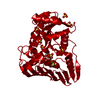
| Title | Crystal structure of oxidoreductase, Gfo/Idh/MocA family from Streptococcus pneumoniae with reductive methylated Lysine | |||||||||
 Components Components | Oxidoreductase, Gfo/Idh/MocA family | |||||||||
 Keywords Keywords | OXIDOREDUCTASE / structural genomics / Midwest Center for Structural Genomics / PSI-Biology / MCSG / Gfo/Idh/MocA family / reductive methylation | |||||||||
| Function / homology |  Function and homology information Function and homology information | |||||||||
| Biological species |   Streptococcus pneumoniae (bacteria) Streptococcus pneumoniae (bacteria) | |||||||||
| Method | X-RAY DIFFRACTION / SYNCHROTRON / SAD / Resolution: 2 Å | |||||||||
 Authors Authors | Chang, C. / Hatzos-Skintges, C. / Abdullah, J. / Joachimiak, A. / Midwest Center for Structural Genomics (MCSG) | |||||||||
 Citation Citation | Journal: TO BE PUBLISHED Title: Crystal structure of oxidoreductase, Gfo/Idh/MocA family from Streptococcus pneumoniae with reductive methylated Lysine Authors: Chang, C. / Hatzos-Skintges, C. / Abdullah, J. / Joachimiak, A. | |||||||||
| History |
|
- Structure visualization
| Structure viewer | Molecule: MolmilJmol/JSmol |
|---|
- Downloads & links
-Download
| PDBx/mmCIF format | 4iq0.cif.gz | 517.7 KB | Display | PDBx/mmCIF format |
|---|---|---|---|---|
| PDB format | pdb4iq0.ent.gz | 441.6 KB | Display | PDB format |
| PDBx/mmJSON format | 4iq0.json.gz | Tree view | PDBx/mmJSON format | |
| Others |  Other downloads Other downloads |
-Validation report
| Arichive directory | https://data.pdbj.org/pub/pdb/validation_reports/iq/4iq0ftp://data.pdbj.org/pub/pdb/validation_reports/iq/4iq0 | HTTPS FTP |
|---|
-Related structure data
| Similar structure data | |
|---|---|
| Other databases |


-Links
 PDBj
PDBj- Assembly
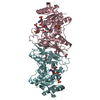
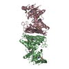
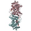
| Deposited unit | 
| ||||||||
|---|---|---|---|---|---|---|---|---|---|
| 1 | 
| ||||||||
| 2 | 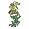
| ||||||||
| Unit cell |
|
-Components
| #1: Protein | Mass: 37018.719 Da / Num. of mol.: 4 Source method: isolated from a genetically manipulated source Source: (gene. exp.) Streptococcus pneumoniae (bacteria) / Gene: SP_1482 / Production host: Escherichia coli (E. coli) / Strain (production host): BL21(DE3)magic / References: UniProt: Q97PV8, UniProt: A0A0H2UR04*PLUS#2: Chemical | ChemComp-K /   Mass: 39.098 Da / Num. of mol.: 5 / Source method: obtained synthetically / Formula: K Mass: 39.098 Da / Num. of mol.: 5 / Source method: obtained synthetically / Formula: K#3: Water | ChemComp-HOH / | Water Mass: 18.015 Da / Num. of mol.: 904 / Source method: isolated from a natural source / Formula: H2O Mass: 18.015 Da / Num. of mol.: 904 / Source method: isolated from a natural source / Formula: H2O |
|---|
-Experimental details
-Experiment
| Experiment | Method: X-RAY DIFFRACTION / Number of used crystals: 1 |
|---|
- Sample preparation
| Crystal | Density Matthews: 2.31 Å3/Da / Density % sol: 46.67 % |
|---|---|
| Crystal grow | Temperature: 289 K / Method: vapor diffusion, sitting drop / pH: 8.5 Details: 0.2 M Lithium sulfate, 0.1 M TRIS, 1.26 M Ammonium sulfate, pH 8.5, VAPOR DIFFUSION, SITTING DROP, temperature 289K |
-Data collection
| Diffraction | Mean temperature: 100 K |
|---|---|
| Diffraction source | Source: SYNCHROTRON / Site: APS  / Beamline: 19-ID / Wavelength: 0.9794 Å / Beamline: 19-ID / Wavelength: 0.9794 Å |
| Detector | Type: ADSC QUANTUM 315r / Detector: CCD / Date: Dec 16, 2012 |
| Radiation | Monochromator: Si(111) double crystal / Protocol: SINGLE WAVELENGTH / Monochromatic (M) / Laue (L): M / Scattering type: x-ray |
| Radiation wavelength | Wavelength: 0.9794 Å / Relative weight: 1 |
| Reflection | Resolution: 2→50 Å / Num. all: 91010 / Num. obs: 90011 / % possible obs: 99.7 % / Observed criterion σ(I): -3 / Redundancy: 3.7 % / Rmerge(I) obs: 0.074 / Net I/σ(I): 25.3 |
| Reflection shell | Resolution: 2→2.07 Å / Redundancy: 3.3 % / Rmerge(I) obs: 0.17 / Mean I/σ(I) obs: 7 / Num. unique all: 8840 / % possible all: 98.6 |
- Processing
| Software |
| |||||||||||||||||||||||||||||||||||||||||||||||||||||||||||||||||||||||||||
|---|---|---|---|---|---|---|---|---|---|---|---|---|---|---|---|---|---|---|---|---|---|---|---|---|---|---|---|---|---|---|---|---|---|---|---|---|---|---|---|---|---|---|---|---|---|---|---|---|---|---|---|---|---|---|---|---|---|---|---|---|---|---|---|---|---|---|---|---|---|---|---|---|---|---|---|---|
| Refinement | Method to determine structure: SAD / Resolution: 2→38.6 Å / Cor.coef. Fo:Fc: 0.959 / Cor.coef. Fo:Fc free: 0.935 / Occupancy max: 1 / Occupancy min: 0.5 / SU B: 6.905 / SU ML: 0.089 / Cross valid method: THROUGHOUT / σ(F): 0 / ESU R Free: 0.141 / Stereochemistry target values: MAXIMUM LIKELIHOOD Details: HYDROGENS HAVE BEEN ADDED IN THE RIDING POSITIONS. U VALUES: REFINED INDIVIDUALLY
| |||||||||||||||||||||||||||||||||||||||||||||||||||||||||||||||||||||||||||
| Solvent computation | Ion probe radii: 0.8 Å / Shrinkage radii: 0.8 Å / VDW probe radii: 1.2 Å / Solvent model: MASK | |||||||||||||||||||||||||||||||||||||||||||||||||||||||||||||||||||||||||||
| Displacement parameters | Biso max: 69.34 Å2 / Biso mean: 17.5895 Å2 / Biso min: 4.84 Å2
| |||||||||||||||||||||||||||||||||||||||||||||||||||||||||||||||||||||||||||
| Refinement step | Cycle: LAST / Resolution: 2→38.6 Å
| |||||||||||||||||||||||||||||||||||||||||||||||||||||||||||||||||||||||||||
| Refine LS restraints |
| |||||||||||||||||||||||||||||||||||||||||||||||||||||||||||||||||||||||||||
| LS refinement shell | Resolution: 1.999→2.051 Å / Total num. of bins used: 20
|
