 Movie
Movie Controller


+ Open data

- Basic information
| Entry | Database: PDB / ID: 2i7p | ||||||
|---|---|---|---|---|---|---|---|
| Title | Crystal structure of human PANK3 in complex with AcCoA | ||||||
 Components Components | Pantothenate kinase 3 | ||||||
 Keywords Keywords | TRANSFERASE / PanK / Structural Genomics / Structural Genomics Consortium / SGC | ||||||
| Function / homology |  Function and homology information Function and homology informationCoenzyme A biosynthesis / vitamin binding / acetyl-CoA binding / pantothenate kinase / pantothenate kinase activity / coenzyme A biosynthetic process / phosphorylation / protein homodimerization activity / ATP binding / nucleus / cytosolSimilarity search - Function | ||||||
| Biological species |  Homo sapiens (human) Homo sapiens (human) | ||||||
| Method | X-RAY DIFFRACTION / SYNCHROTRON / MOLECULAR REPLACEMENT / Resolution: 2.05 Å | ||||||
 Authors Authors | Hong, B.S. / Wang, L. / Shen, L. / Tempel, W. / Loppnau, P. / Finerty, P. / Arrowsmith, C.H. / Edwards, A.M. / Sundstrom, M. / Weigelt, J. ...Hong, B.S. / Wang, L. / Shen, L. / Tempel, W. / Loppnau, P. / Finerty, P. / Arrowsmith, C.H. / Edwards, A.M. / Sundstrom, M. / Weigelt, J. / Bochkarev, A. / Park, H.W. / Structural Genomics Consortium (SGC) | ||||||
 Citation Citation | Journal: J.Biol.Chem. / Year: 2007 Title: Crystal structures of human pantothenate kinases. Insights into allosteric regulation and mutations linked to a neurodegeneration disorder. Authors: Hong, B.S. / Senisterra, G. / Rabeh, W.M. / Vedadi, M. / Leonardi, R. / Zhang, Y.M. / Rock, C.O. / Jackowski, S. / Park, H.W. | ||||||
| History |
|
- Structure visualization
| Structure viewer | Molecule: MolmilJmol/JSmol |
|---|
- Downloads & links
-Download
| PDBx/mmCIF format | 2i7p.cif.gz | 296.8 KB | Display | PDBx/mmCIF format |
|---|---|---|---|---|
| PDB format | pdb2i7p.ent.gz | 238.1 KB | Display | PDB format |
| PDBx/mmJSON format | 2i7p.json.gz | Tree view | PDBx/mmJSON format | |
| Others |  Other downloads Other downloads |
-Validation report
| Arichive directory | https://data.pdbj.org/pub/pdb/validation_reports/i7/2i7pftp://data.pdbj.org/pub/pdb/validation_reports/i7/2i7p | HTTPS FTP |
|---|
-Related structure data


-Links
 PDBj
PDBj- Assembly
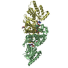
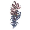
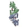
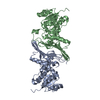
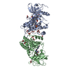
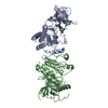
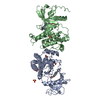
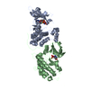
| Deposited unit | 
| ||||||||
|---|---|---|---|---|---|---|---|---|---|
| 1 | 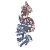
| ||||||||
| 2 | 
| ||||||||
| Unit cell |
| ||||||||
| Details | The biological assembly is a dimer, and two dimers in the asymmetric unit |
-Components
| #1: Protein | / Pantothenic acid kinase 3 / hPanK3 Mass: 40125.688 Da / Num. of mol.: 4 Source method: isolated from a genetically manipulated source Source: (gene. exp.) Homo sapiens (human) / Gene: PANK3 / Plasmid: pET28aLIC / Species (production host): Escherichia coli / Production host:  Escherichia coli BL21(DE3) (bacteria) / Strain (production host): BL21(DE3) / References: UniProt: Q9H999, pantothenate kinase Escherichia coli BL21(DE3) (bacteria) / Strain (production host): BL21(DE3) / References: UniProt: Q9H999, pantothenate kinase#2: Chemical | ChemComp-ACO / Acetyl-CoA  Mass: 809.571 Da / Num. of mol.: 4 / Source method: obtained synthetically / Formula: C23H38N7O17P3S Mass: 809.571 Da / Num. of mol.: 4 / Source method: obtained synthetically / Formula: C23H38N7O17P3S#3: Water | ChemComp-HOH / | Water Mass: 18.015 Da / Num. of mol.: 715 / Source method: isolated from a natural source / Formula: H2O Mass: 18.015 Da / Num. of mol.: 715 / Source method: isolated from a natural source / Formula: H2O |
|---|
-Experimental details
-Experiment
| Experiment | Method: X-RAY DIFFRACTION / Number of used crystals: 1 |
|---|
- Sample preparation
| Crystal | Density Matthews: 2.46 Å3/Da / Density % sol: 50.05 % |
|---|---|
| Crystal grow | Temperature: 298 K / Method: vapor diffusion, sitting drop / pH: 8.5 Details: 1M Tris, 50% PEG3350, 2M NH4Citrate, pH 8.5, VAPOR DIFFUSION, SITTING DROP, temperature 298K |
-Data collection
| Diffraction | Mean temperature: 100 K |
|---|---|
| Diffraction source | Source: SYNCHROTRON / Site: APS  / Beamline: 19-BM / Wavelength: 0.9 Å / Beamline: 19-BM / Wavelength: 0.9 Å |
| Detector | Type: SBC-3 / Detector: CCD / Date: Feb 25, 2006 |
| Radiation | Monochromator: Rosenbaum-Rock double-crystal monochromator / Protocol: SINGLE WAVELENGTH / Monochromatic (M) / Laue (L): M / Scattering type: x-ray |
| Radiation wavelength | Wavelength: 0.9 Å / Relative weight: 1 |
| Reflection | Resolution: 2.05→30 Å / Num. obs: 96048 / % possible obs: 98.8 % / Observed criterion σ(F): 1 / Observed criterion σ(I): 1 / Redundancy: 3.5 % / Biso Wilson estimate: 22.7 Å2 / Rmerge(I) obs: 0.061 / Rsym value: 0.088 / Net I/σ(I): 13.3 |
| Reflection shell | Resolution: 2.05→2.12 Å / Redundancy: 2.3 % / Rmerge(I) obs: 0.446 / Mean I/σ(I) obs: 1.27 / Num. unique all: 8777 / Rsym value: 0.522 / % possible all: 90.5 |
- Processing
| Software |
| ||||||||||||||||||||||||||||||||||||||||||||||||||||||||||||
|---|---|---|---|---|---|---|---|---|---|---|---|---|---|---|---|---|---|---|---|---|---|---|---|---|---|---|---|---|---|---|---|---|---|---|---|---|---|---|---|---|---|---|---|---|---|---|---|---|---|---|---|---|---|---|---|---|---|---|---|---|---|
| Refinement | Method to determine structure: MOLECULAR REPLACEMENT / Resolution: 2.05→29.34 Å / Rfactor Rfree error: 0.004 / Data cutoff high absF: 2434781.55 / Data cutoff low absF: 0 / Isotropic thermal model: RESTRAINED / Cross valid method: THROUGHOUT / σ(F): 0 / Details: HYDROGENS HAVE BEEN ADDED IN THE RIDING POSITIONS
| ||||||||||||||||||||||||||||||||||||||||||||||||||||||||||||
| Solvent computation | Solvent model: FLAT MODEL / Bsol: 42.2418 Å2 / ksol: 0.346971 e/Å3 | ||||||||||||||||||||||||||||||||||||||||||||||||||||||||||||
| Displacement parameters | Biso mean: 32.7 Å2
| ||||||||||||||||||||||||||||||||||||||||||||||||||||||||||||
| Refine analyze |
| ||||||||||||||||||||||||||||||||||||||||||||||||||||||||||||
| Refinement step | Cycle: LAST / Resolution: 2.05→29.34 Å
| ||||||||||||||||||||||||||||||||||||||||||||||||||||||||||||
| Refine LS restraints |
| ||||||||||||||||||||||||||||||||||||||||||||||||||||||||||||
| LS refinement shell | Resolution: 1.9→2.02 Å / Total num. of bins used: 6 /
| ||||||||||||||||||||||||||||||||||||||||||||||||||||||||||||
| Xplor file |
|
