 Movie
Movie Controller

[English] 日本語
 Yorodumi
- PDB-2ht2: Structure of the Escherichia coli ClC chloride channel Y445H muta... -
+ Open data

- Basic information
| Entry | Database: PDB / ID: 2ht2 | ||||||
|---|---|---|---|---|---|---|---|
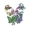
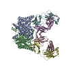
| Title | Structure of the Escherichia coli ClC chloride channel Y445H mutant and Fab complex | ||||||
 Components Components |
| ||||||
 Keywords Keywords |  MEMBRANE PROTEIN / ClC family of channel and transporters / H+/Cl- antiporter / Fab complex MEMBRANE PROTEIN / ClC family of channel and transporters / H+/Cl- antiporter / Fab complex | ||||||
| Function / homology |  Function and homology information Function and homology informationchloride:proton antiporter activity / cellular stress response to acidic pH / voltage-gated chloride channel activity / chloride transmembrane transport / proton transmembrane transport / identical protein binding / plasma membraneSimilarity search - Function | ||||||
| Biological species |  Escherichia coli (E. coli) Escherichia coli (E. coli) Mus musculus (house mouse) Mus musculus (house mouse) | ||||||
| Method | X-RAY DIFFRACTION / SYNCHROTRON / MOLECULAR REPLACEMENT / Resolution: 3.32 Å | ||||||
 Authors Authors | Accardi, A. / Lobet, S. / Williams, C. / Miller, C. / Dutzler, R. | ||||||
 Citation Citation | Journal: J.Mol.Biol. / Year: 2006 Title: Synergism Between Halide Binding and Proton Transport in a CLC-type Exchanger. Authors: Accardi, A. / Lobet, S. / Williams, C. / Miller, C. / Dutzler, R. | ||||||
| History |
| ||||||
| Remark 600 | HETEROGEN EACH SUBUNIT OF THE MUTANT HAS A SINGLE BR- ION BOUND IN THE SELECTIVITY FILTER |
- Structure visualization
| Structure viewer | Molecule: MolmilJmol/JSmol |
|---|
- Downloads & links
-Download
| PDBx/mmCIF format | 2ht2.cif.gz | 331.2 KB | Display | PDBx/mmCIF format |
|---|---|---|---|---|
| PDB format | pdb2ht2.ent.gz | 276.6 KB | Display | PDB format |
| PDBx/mmJSON format | 2ht2.json.gz | Tree view | PDBx/mmJSON format | |
| Others |  Other downloads Other downloads |
-Validation report
| Arichive directory | https://data.pdbj.org/pub/pdb/validation_reports/ht/2ht2ftp://data.pdbj.org/pub/pdb/validation_reports/ht/2ht2 | HTTPS FTP |
|---|
-Related structure data


-Links
 PDBj
PDBj


- Assembly
| Deposited unit | 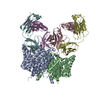
| ||||||||||||||||||
|---|---|---|---|---|---|---|---|---|---|---|---|---|---|---|---|---|---|---|---|
| 1 |
| ||||||||||||||||||
| Unit cell |
| ||||||||||||||||||
| Noncrystallographic symmetry (NCS) | NCS domain:
NCS domain segments: Component-ID: 1 / Ens-ID: 1 / Beg label comp-ID: ARG / End label comp-ID: ALA / Refine code: 5 / Auth seq-ID: 18 - 458 / Label seq-ID: 18 - 458
|
-Components
| #1: Protein | Mass: 50365.375 Da / Num. of mol.: 2 / Mutation: Y445H Source method: isolated from a genetically manipulated source Source: (gene. exp.) Escherichia coli (E. coli) / Gene: clcA, eriC / Plasmid: pet 28b+ / Species (production host): Escherichia coli / Production host: Escherichia coli BL21(DE3) (bacteria) / Strain (production host): BL21DE3 / References: UniProt: P37019#2: Antibody | Fragment antigen-bindingMass: 23693.918 Da / Num. of mol.: 2 / Source method: isolated from a natural source / Source: (natural) Mus musculus (house mouse) / Cell line: HYBRIDOMA CELL LINE / References: UniProt: Q4VBH1*PLUS#3: Antibody | Fragment antigen-bindingMass: 23088.443 Da / Num. of mol.: 2 / Source method: isolated from a natural source / Source: (natural) Mus musculus (house mouse) / Cell line: HYBRIDOMA CELL LINE#4: Chemical | Bromide  Mass: 79.904 Da / Num. of mol.: 2 / Source method: obtained synthetically / Formula: Br Mass: 79.904 Da / Num. of mol.: 2 / Source method: obtained synthetically / Formula: Br |
|---|
-Experimental details
-Experiment
| Experiment | Method: X-RAY DIFFRACTION / Number of used crystals: 1 |
|---|
- Sample preparation
| Crystal | Density Matthews: 3.81 Å3/Da / Density % sol: 67.7 % |
|---|---|
| Crystal grow | Temperature: 293 K / Method: vapor diffusion, sitting drop / pH: 8.5 Details: 38% peg 300, 50mM Tris, 150 mM NaKTart, pH 8.5, VAPOR DIFFUSION, SITTING DROP, temperature 293K |
-Data collection
| Diffraction | Mean temperature: 100 K |
|---|---|
| Diffraction source | Source: SYNCHROTRON / Site: ALS  / Beamline: 8.2.1 / Wavelength: 0.9193 Å / Beamline: 8.2.1 / Wavelength: 0.9193 Å |
| Detector | Type: ADSC QUANTUM 210 / Detector: CCD / Date: Jan 14, 2006 |
| Radiation | Monochromator: Double crystal, Si(111) / Protocol: SINGLE WAVELENGTH / Monochromatic (M) / Laue (L): M / Scattering type: x-ray |
| Radiation wavelength | Wavelength: 0.9193 Å / Relative weight: 1 |
| Reflection | Resolution: 3.32→40 Å / Num. all: 39928 / Num. obs: 37472 / % possible obs: 93.85 % / Observed criterion σ(F): 1 / Observed criterion σ(I): 1 / Biso Wilson estimate: 87.552 Å2 / Rmerge(I) obs: 0.089 |
| Reflection shell | Resolution: 3.32→3.41 Å / % possible all: 94.83 |
- Processing
| Software |
| ||||||||||||||||||||||||||||||||||||||||||||||||||||||||||||||||||||||||||||||||||||||||||
|---|---|---|---|---|---|---|---|---|---|---|---|---|---|---|---|---|---|---|---|---|---|---|---|---|---|---|---|---|---|---|---|---|---|---|---|---|---|---|---|---|---|---|---|---|---|---|---|---|---|---|---|---|---|---|---|---|---|---|---|---|---|---|---|---|---|---|---|---|---|---|---|---|---|---|---|---|---|---|---|---|---|---|---|---|---|---|---|---|---|---|---|
| Refinement | Method to determine structure: MOLECULAR REPLACEMENT / Resolution: 3.32→40 Å / Cor.coef. Fo:Fc: 0.905 / Cor.coef. Fo:Fc free: 0.889 / Cross valid method: THROUGHOUT / ESU R: 0.558 / ESU R Free: 0.626 / Stereochemistry target values: MAXIMUM LIKELIHOOD / Details: HYDROGENS HAVE BEEN ADDED IN THE RIDING POSITIONS
| ||||||||||||||||||||||||||||||||||||||||||||||||||||||||||||||||||||||||||||||||||||||||||
| Solvent computation | Ion probe radii: 0.8 Å / Shrinkage radii: 0.8 Å / VDW probe radii: 1.2 Å / Solvent model: MASK | ||||||||||||||||||||||||||||||||||||||||||||||||||||||||||||||||||||||||||||||||||||||||||
| Displacement parameters | Biso mean: 86.957 Å2
| ||||||||||||||||||||||||||||||||||||||||||||||||||||||||||||||||||||||||||||||||||||||||||
| Refinement step | Cycle: LAST / Resolution: 3.32→40 Å
| ||||||||||||||||||||||||||||||||||||||||||||||||||||||||||||||||||||||||||||||||||||||||||
| Refine LS restraints |
| ||||||||||||||||||||||||||||||||||||||||||||||||||||||||||||||||||||||||||||||||||||||||||
| Refine LS restraints NCS | Dom-ID: 1 / Auth asym-ID: A / Ens-ID: 1 / Refine-ID: X-RAY DIFFRACTION
| ||||||||||||||||||||||||||||||||||||||||||||||||||||||||||||||||||||||||||||||||||||||||||
| LS refinement shell | Resolution: 3.32→3.406 Å / Total num. of bins used: 20
|
