 Movie
Movie Controller

[English] 日本語
 Yorodumi
- PDB-3det: Structure of the E148A, Y445A doubly ungated mutant of E.coli CLC... -
+ Open data

- Basic information
| Entry | Database: PDB / ID: 3det | ||||||
|---|---|---|---|---|---|---|---|
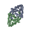
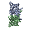
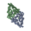
| Title | Structure of the E148A, Y445A doubly ungated mutant of E.coli CLC_Ec1, Cl-/H+ antiporter | ||||||
 Components Components |
| ||||||
 Keywords Keywords |  MEMBRANE PROTEIN / CLC_Ec1 / antiporter / exchange-transporter / doubly ungated mutant / Chloride / Inner membrane / Ion transport / Membrane / Stress response / Transmembrane MEMBRANE PROTEIN / CLC_Ec1 / antiporter / exchange-transporter / doubly ungated mutant / Chloride / Inner membrane / Ion transport / Membrane / Stress response / Transmembrane | ||||||
| Function / homology |  Function and homology information Function and homology informationchloride:proton antiporter activity / cellular stress response to acidic pH / voltage-gated chloride channel activity / chloride transmembrane transport / proton transmembrane transport / identical protein binding / plasma membraneSimilarity search - Function | ||||||
| Biological species |  Escherichia coli (E. coli) Escherichia coli (E. coli) Mus musculus (house mouse) Mus musculus (house mouse) | ||||||
| Method | X-RAY DIFFRACTION / SYNCHROTRON / MOLECULAR REPLACEMENT / Resolution: 2.8 Å | ||||||
 Authors Authors | Jayaram, H. / Accardi, A. / Wu, F. / Williams, C. / Miller, C. | ||||||
 Citation Citation | Journal: Proc.Natl.Acad.Sci.USA / Year: 2008 Title: Ion permeation through a Cl--selective channel designed from a CLC Cl-/H+ exchanger Authors: Jayaram, H. / Accardi, A. / Wu, F. / Williams, C. / Miller, C. | ||||||
| History |
|
- Structure visualization
| Structure viewer | Molecule: MolmilJmol/JSmol |
|---|
- Downloads & links
-Download
| PDBx/mmCIF format | 3det.cif.gz | 328.7 KB | Display | PDBx/mmCIF format |
|---|---|---|---|---|
| PDB format | pdb3det.ent.gz | 267.7 KB | Display | PDB format |
| PDBx/mmJSON format | 3det.json.gz | Tree view | PDBx/mmJSON format | |
| Others |  Other downloads Other downloads |
-Validation report
| Arichive directory | https://data.pdbj.org/pub/pdb/validation_reports/de/3detftp://data.pdbj.org/pub/pdb/validation_reports/de/3det | HTTPS FTP |
|---|
-Related structure data
| Related structure data | 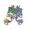 1otsS S: Starting model for refinement |
|---|---|
| Similar structure data |


-Links
 PDBj
PDBj

- Assembly
| Deposited unit | 
| ||||||||
|---|---|---|---|---|---|---|---|---|---|
| 1 | 
| ||||||||
| 2 |
| ||||||||
| Unit cell |
|
-Components
| #1: Protein | Mass: 50240.273 Da / Num. of mol.: 2 / Mutation: E148A, Y445A Source method: isolated from a genetically manipulated source Source: (gene. exp.) Escherichia coli (E. coli) / Strain: K12 / Gene: clcA, eriC, yadQ / Plasmid: pASK / Production host: Escherichia coli (E. coli) / Strain (production host): Bl21(DE3) / References: UniProt: P37019#2: Antibody | Fragment antigen-bindingMass: 23693.918 Da / Num. of mol.: 2 / Source method: isolated from a natural source / Source: (natural) Mus musculus (house mouse)#3: Antibody | Fragment antigen-bindingMass: 23088.443 Da / Num. of mol.: 2 / Source method: isolated from a natural source / Source: (natural) Mus musculus (house mouse) |
|---|
-Experimental details
-Experiment
| Experiment | Method: X-RAY DIFFRACTION / Number of used crystals: 1 |
|---|
- Sample preparation
| Crystal | Density Matthews: 3.69 Å3/Da / Density % sol: 66.64 % |
|---|---|
| Crystal grow | Temperature: 298 K / Method: vapor diffusion, sitting drop / pH: 5.5 Details: 300 mM KCl, 26.6% PEG 600, 50 mM Na-cacodylate pH 5.5, VAPOR DIFFUSION, SITTING DROP, temperature 298K |
-Data collection
| Diffraction | Mean temperature: 178 K |
|---|---|
| Diffraction source | Source: SYNCHROTRON / Site: APS  / Beamline: 23-ID-D / Wavelength: 0.97949 Å / Beamline: 23-ID-D / Wavelength: 0.97949 Å |
| Detector | Type: MARMOSAIC 300 mm CCD / Detector: CCD / Date: Apr 19, 2008 |
| Radiation | Protocol: SINGLE WAVELENGTH / Monochromatic (M) / Laue (L): M / Scattering type: x-ray |
| Radiation wavelength | Wavelength: 0.97949 Å / Relative weight: 1 |
| Reflection | Resolution: 2.8→86 Å / Num. all: 69549 / Num. obs: 60533 / % possible obs: 99.9 % / Observed criterion σ(F): 2 / Observed criterion σ(I): 2 / Redundancy: 7.3 % / Rmerge(I) obs: 0.106 / Rsym value: 0.106 / Net I/σ(I): 10.9 |
| Reflection shell | Resolution: 2.8→2.86 Å / Redundancy: 6.9 % / Rmerge(I) obs: 0.16 / Mean I/σ(I) obs: 4.2 / Num. unique all: 10078 / Rsym value: 0.16 / % possible all: 99.8 |
- Processing
| Software |
| |||||||||||||||||||||||||||||||||||||||||||||||||||||||||||||||||
|---|---|---|---|---|---|---|---|---|---|---|---|---|---|---|---|---|---|---|---|---|---|---|---|---|---|---|---|---|---|---|---|---|---|---|---|---|---|---|---|---|---|---|---|---|---|---|---|---|---|---|---|---|---|---|---|---|---|---|---|---|---|---|---|---|---|---|
| Refinement | Method to determine structure: MOLECULAR REPLACEMENT Starting model: PDB entry 1OTS Resolution: 2.8→59.03 Å / Cor.coef. Fo:Fc: 0.919 / Cor.coef. Fo:Fc free: 0.892 / SU B: 20.381 / SU ML: 0.378 / Cross valid method: THROUGHOUT- 1OTS structure factor file / σ(F): 0 / ESU R: 0.977 / ESU R Free: 0.41 / Stereochemistry target values: MAXIMUM LIKELIHOOD
| |||||||||||||||||||||||||||||||||||||||||||||||||||||||||||||||||
| Solvent computation | Ion probe radii: 0.8 Å / Shrinkage radii: 0.8 Å / VDW probe radii: 1.2 Å / Solvent model: BABINET MODEL WITH MASK | |||||||||||||||||||||||||||||||||||||||||||||||||||||||||||||||||
| Displacement parameters | Biso mean: 101.565 Å2
| |||||||||||||||||||||||||||||||||||||||||||||||||||||||||||||||||
| Refinement step | Cycle: LAST / Resolution: 2.8→59.03 Å
| |||||||||||||||||||||||||||||||||||||||||||||||||||||||||||||||||
| Refine LS restraints |
| |||||||||||||||||||||||||||||||||||||||||||||||||||||||||||||||||
| LS refinement shell | Resolution: 2.8→2.872 Å / Total num. of bins used: 20
|
