 Movie
Movie Controller


+ Open data

- Basic information
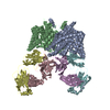
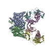
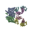
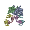
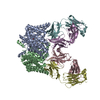
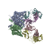
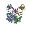
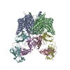
| Entry | Database: PDB / ID: 2r9h | ||||||
|---|---|---|---|---|---|---|---|
| Title | Crystal Structure of Q207C Mutant of CLC-ec1 in complex with Fab | ||||||
 Components Components |
| ||||||
 Keywords Keywords |  MEMBRANE PROTEIN / CLC / antiporter / transporter / exchanger / disulfide / crosslink / Chloride / Inner membrane / Ion transport / Stress response / Transmembrane MEMBRANE PROTEIN / CLC / antiporter / transporter / exchanger / disulfide / crosslink / Chloride / Inner membrane / Ion transport / Stress response / Transmembrane | ||||||
| Function / homology |  Function and homology information Function and homology informationchloride:proton antiporter activity / cellular stress response to acidic pH / voltage-gated chloride channel activity / chloride transmembrane transport / proton transmembrane transport / identical protein binding / plasma membraneSimilarity search - Function | ||||||
| Biological species |  Escherichia coli (E. coli) Escherichia coli (E. coli) Mus musculus (house mouse) Mus musculus (house mouse) | ||||||
| Method | X-RAY DIFFRACTION / SYNCHROTRON / MOLECULAR REPLACEMENT / molecular replacement / Resolution: 3.1 Å | ||||||
 Authors Authors | Nguitragool, W. / Miller, C. | ||||||
 Citation Citation | Journal: To be Published Title: Crystal Structure of Q207C Mutant of CLC-ec1 in complex with Fab Authors: Miller, C. / Nguitragool, W. | ||||||
| History |
|
- Structure visualization
| Structure viewer | Molecule: MolmilJmol/JSmol |
|---|
- Downloads & links
-Download
| PDBx/mmCIF format | 2r9h.cif.gz | 328.3 KB | Display | PDBx/mmCIF format |
|---|---|---|---|---|
| PDB format | pdb2r9h.ent.gz | 273.1 KB | Display | PDB format |
| PDBx/mmJSON format | 2r9h.json.gz | Tree view | PDBx/mmJSON format | |
| Others |  Other downloads Other downloads |
-Validation report
| Arichive directory | https://data.pdbj.org/pub/pdb/validation_reports/r9/2r9hftp://data.pdbj.org/pub/pdb/validation_reports/r9/2r9h | HTTPS FTP |
|---|
-Related structure data
| Similar structure data |
|---|


-Links
 PDBj
PDBj

- Assembly
| Deposited unit | 
| ||||||||||||||||||
|---|---|---|---|---|---|---|---|---|---|---|---|---|---|---|---|---|---|---|---|
| 1 |
| ||||||||||||||||||
| Unit cell |
| ||||||||||||||||||
| Noncrystallographic symmetry (NCS) | NCS domain:
NCS domain segments: Component-ID: 1 / Ens-ID: 1 / Beg label comp-ID: ARG / End label comp-ID: ALA / Refine code: 3 / Auth seq-ID: 18 - 458 / Label seq-ID: 2 - 442
|
-Components
| #1: Protein | Mass: 47333.043 Da / Num. of mol.: 2 / Mutation: Q207C Source method: isolated from a genetically manipulated source Source: (gene. exp.) Escherichia coli (E. coli) / Gene: clcA, eriC / Plasmid: pASK90 / Production host: Escherichia coli (E. coli) / Strain (production host): BL21 / References: UniProt: P37019#2: Antibody | Fragment antigen-bindingMass: 23693.918 Da / Num. of mol.: 2 / Source method: isolated from a natural source / Source: (natural) Mus musculus (house mouse)#3: Antibody | Fragment antigen-bindingMass: 23088.443 Da / Num. of mol.: 2 / Source method: isolated from a natural source / Source: (natural) Mus musculus (house mouse)#4: Chemical | ChemComp-CL / Chloride  Mass: 35.453 Da / Num. of mol.: 4 / Source method: obtained synthetically / Formula: Cl Mass: 35.453 Da / Num. of mol.: 4 / Source method: obtained synthetically / Formula: Cl |
|---|
-Experimental details
-Experiment
| Experiment | Method: X-RAY DIFFRACTION / Number of used crystals: 1 |
|---|
- Sample preparation
| Crystal | Density Matthews: 3.9 Å3/Da / Density % sol: 68.5 % |
|---|---|
| Crystal grow | Temperature: 295 K / Method: sitting drop / pH: 9.5 Details: Vapor diffusion: 3 uL of protein complex, 7 mg/ml, in 50 mM NaKTartrate, 5 mM Tris, 12.5 mM NaCl, 18.5% w/v PEG300, 25 mM glycine against 300 uL of 25 mM NaCl, 37% w/v PEG300, 50 mM glycine, ...Details: Vapor diffusion: 3 uL of protein complex, 7 mg/ml, in 50 mM NaKTartrate, 5 mM Tris, 12.5 mM NaCl, 18.5% w/v PEG300, 25 mM glycine against 300 uL of 25 mM NaCl, 37% w/v PEG300, 50 mM glycine, pH 9.5, sitting drop, temperature 295K |
-Data collection
| Diffraction source | Source: SYNCHROTRON / Site: NSLS  / Beamline: X29A / Wavelength: 0.9204 Å / Beamline: X29A / Wavelength: 0.9204 Å | |||||||||||||||||||||||||||||||||||||||||||||||||||||||||||||||||||||||||||||
|---|---|---|---|---|---|---|---|---|---|---|---|---|---|---|---|---|---|---|---|---|---|---|---|---|---|---|---|---|---|---|---|---|---|---|---|---|---|---|---|---|---|---|---|---|---|---|---|---|---|---|---|---|---|---|---|---|---|---|---|---|---|---|---|---|---|---|---|---|---|---|---|---|---|---|---|---|---|---|
| Detector | Type: ADSC QUANTUM 315 / Detector: CCD | |||||||||||||||||||||||||||||||||||||||||||||||||||||||||||||||||||||||||||||
| Radiation | Protocol: SINGLE WAVELENGTH / Monochromatic (M) / Laue (L): M / Scattering type: x-ray | |||||||||||||||||||||||||||||||||||||||||||||||||||||||||||||||||||||||||||||
| Radiation wavelength | Wavelength: 0.9204 Å / Relative weight: 1 | |||||||||||||||||||||||||||||||||||||||||||||||||||||||||||||||||||||||||||||
| Reflection | Resolution: 3.1→50 Å / Num. obs: 52974 / % possible obs: 99.2 % / Redundancy: 6.7 % / Rmerge(I) obs: 0.08 / Χ2: 2.227 / Net I/σ(I): 14.4 | |||||||||||||||||||||||||||||||||||||||||||||||||||||||||||||||||||||||||||||
| Reflection shell |
|
-Phasing
| Phasing | Method: molecular replacement | |||||||||
|---|---|---|---|---|---|---|---|---|---|---|
| Phasing MR | Model details: Phaser MODE: MR_AUTO
|
- Processing
| Software |
| ||||||||||||||||||||||||||||||||||||||||||||||||||||||||||||||||||||||
|---|---|---|---|---|---|---|---|---|---|---|---|---|---|---|---|---|---|---|---|---|---|---|---|---|---|---|---|---|---|---|---|---|---|---|---|---|---|---|---|---|---|---|---|---|---|---|---|---|---|---|---|---|---|---|---|---|---|---|---|---|---|---|---|---|---|---|---|---|---|---|---|
| Refinement | Method to determine structure: MOLECULAR REPLACEMENT / Resolution: 3.1→50 Å / Cor.coef. Fo:Fc: 0.909 / Cor.coef. Fo:Fc free: 0.908 / Cross valid method: THROUGHOUT / σ(F): 0 / ESU R: 0.913 / ESU R Free: 0.454 / Stereochemistry target values: MAXIMUM LIKELIHOOD / Details: HYDROGENS HAVE BEEN ADDED IN THE RIDING POSITIONS
| ||||||||||||||||||||||||||||||||||||||||||||||||||||||||||||||||||||||
| Solvent computation | Ion probe radii: 0.8 Å / Shrinkage radii: 0.8 Å / VDW probe radii: 1.4 Å / Solvent model: MASK | ||||||||||||||||||||||||||||||||||||||||||||||||||||||||||||||||||||||
| Displacement parameters | Biso mean: 105.226 Å2
| ||||||||||||||||||||||||||||||||||||||||||||||||||||||||||||||||||||||
| Refinement step | Cycle: LAST / Resolution: 3.1→50 Å
| ||||||||||||||||||||||||||||||||||||||||||||||||||||||||||||||||||||||
| Refine LS restraints |
| ||||||||||||||||||||||||||||||||||||||||||||||||||||||||||||||||||||||
| Refine LS restraints NCS | Dom-ID: 1 / Auth asym-ID: A / Ens-ID: 1 / Refine-ID: X-RAY DIFFRACTION
| ||||||||||||||||||||||||||||||||||||||||||||||||||||||||||||||||||||||
| LS refinement shell | Resolution: 3.1→3.18 Å / Total num. of bins used: 20
|
