 Movie
Movie Controller

[English] 日本語
 Yorodumi
- PDB-2ek9: Aminopeptidase from Aneurinibacillus sp. strain AM-1 with Bestatin -
+ Open data

- Basic information
| Entry | Database: PDB / ID: 2ek9 | ||||||
|---|---|---|---|---|---|---|---|
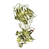
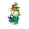
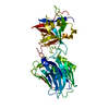
| Title | Aminopeptidase from Aneurinibacillus sp. strain AM-1 with Bestatin | ||||||
 Components Components | Aminopeptidase | ||||||
 Keywords Keywords | HYDROLASE / METALLOPROTEINASE | ||||||
| Function / homology |  Function and homology informationmetalloexopeptidase activity / aminopeptidase activity / proteolysis / metal ion binding Function and homology informationmetalloexopeptidase activity / aminopeptidase activity / proteolysis / metal ion bindingSimilarity search - Function | ||||||
| Biological species |  Aneurinibacillus sp. (bacteria) Aneurinibacillus sp. (bacteria) | ||||||
| Method | X-RAY DIFFRACTION / MOLECULAR REPLACEMENT / Resolution: 1.97 Å | ||||||
 Authors Authors | Akioka, M. / Nakano, H. / Watanabe, K. | ||||||
 Citation Citation | Journal: To be published Title: Structural characterization of a novel bacterial aminopeptidase with an apical domain from aneurinibacillus sp. strain AM-1 Authors: Akioka, M. / Nakano, H. / Tsujimoto, Y. / Matsui, H. / Nakatsu, T. / Kato, H. / Watanabe, K. #1: Journal: ACTA CRYSTALLOGR.,SECT.F / Year: 2006 Title: Overexpression, purification, crystallization and preliminary X-ray crystallographic studies of a proline-specific aminopeptidase from Aneurinibacillus sp. strain AM-1 Authors: Akioka, M. / Nakano, H. / Horikiri, A. / Tsujimoto, Y. / Matsui, H. / Shimizu, T. / Nakatsu, T. / Kato, H. / Watanabe, K. | ||||||
| History |
|
- Structure visualization
| Structure viewer | Molecule: MolmilJmol/JSmol |
|---|
- Downloads & links
-Download
| PDBx/mmCIF format | 2ek9.cif.gz | 104.9 KB | Display | PDBx/mmCIF format |
|---|---|---|---|---|
| PDB format | pdb2ek9.ent.gz | 77.6 KB | Display | PDB format |
| PDBx/mmJSON format | 2ek9.json.gz | Tree view | PDBx/mmJSON format | |
| Others |  Other downloads Other downloads |
-Validation report
| Arichive directory | https://data.pdbj.org/pub/pdb/validation_reports/ek/2ek9ftp://data.pdbj.org/pub/pdb/validation_reports/ek/2ek9 | HTTPS FTP |
|---|
-Related structure data
| Related structure data | 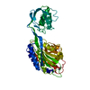 2ek8SC S: Starting model for refinement C: citing same article ( |
|---|---|
| Similar structure data |


-Links
 PDBj
PDBj

- Assembly
| Deposited unit | 
| ||||||||||||||||||
|---|---|---|---|---|---|---|---|---|---|---|---|---|---|---|---|---|---|---|---|
| 1 |
| ||||||||||||||||||
| 2 | 
| ||||||||||||||||||
| Unit cell |
| ||||||||||||||||||
| Components on special symmetry positions |
|
-Components
| #1: Protein | Mass: 45289.699 Da / Num. of mol.: 1 / Fragment: Residues 1-421 Source method: isolated from a genetically manipulated source Source: (gene. exp.) Aneurinibacillus sp. (bacteria) / Strain: AM-1 / Plasmid: PET-11A / Production host: Escherichia coli (E. coli) / References: UniProt: A2V759 | ||||||
|---|---|---|---|---|---|---|---|
| #2: Chemical | ChemComp-ZN /   Mass: 65.409 Da / Num. of mol.: 6 / Source method: obtained synthetically / Formula: Zn Mass: 65.409 Da / Num. of mol.: 6 / Source method: obtained synthetically / Formula: Zn#3: Chemical | ChemComp-BES / | Ubenimex  Mass: 308.373 Da / Num. of mol.: 1 / Source method: obtained synthetically / Formula: C16H24N2O4 / Comment: protease inhibitor*YM Mass: 308.373 Da / Num. of mol.: 1 / Source method: obtained synthetically / Formula: C16H24N2O4 / Comment: protease inhibitor*YM#4: Chemical | ChemComp-IPA / | Isopropyl alcohol  Mass: 60.095 Da / Num. of mol.: 1 / Source method: obtained synthetically / Formula: C3H8O / Comment: alkaloid*YM Mass: 60.095 Da / Num. of mol.: 1 / Source method: obtained synthetically / Formula: C3H8O / Comment: alkaloid*YM#5: Water | ChemComp-HOH / | Water Mass: 18.015 Da / Num. of mol.: 504 / Source method: isolated from a natural source / Formula: H2O Mass: 18.015 Da / Num. of mol.: 504 / Source method: isolated from a natural source / Formula: H2O |
-Experimental details
-Experiment
| Experiment | Method: X-RAY DIFFRACTION / Number of used crystals: 1 |
|---|
- Sample preparation
| Crystal | Density Matthews: 2.74 Å3/Da / Density % sol: 55.09 % |
|---|---|
| Crystal grow | Temperature: 299 K / Method: vapor diffusion / pH: 5.8 Details: 13% PEG 8000, 0.1M MES-NaOH, 0.2M Zinc acetate, pH 5.8, VAPOR DIFFUSION, temperature 299K |
-Data collection
| Diffraction | Mean temperature: 93 K |
|---|---|
| Diffraction source | Source: ROTATING ANODE / Type: RIGAKU / Wavelength: 1.5418 Å |
| Detector | Type: RIGAKU RAXIS VII / Detector: IMAGE PLATE / Date: Aug 29, 2006 |
| Radiation | Protocol: SINGLE WAVELENGTH / Monochromatic (M) / Laue (L): M / Scattering type: x-ray |
| Radiation wavelength | Wavelength: 1.5418 Å / Relative weight: 1 |
| Reflection | Resolution: 1.97→15 Å / Num. obs: 35820 / % possible obs: 99.8 % / Redundancy: 4.75 % / Biso Wilson estimate: 17.3 Å2 / Rmerge(I) obs: 0.069 / Net I/σ(I): 18.9 |
| Reflection shell | Resolution: 1.97→2.08 Å / Rmerge(I) obs: 0.292 / Mean I/σ(I) obs: 4.9 / % possible all: 100 |
- Processing
| Software |
| ||||||||||||||||||||||||||||||||||||||||||||||||||||||||||||||||||||||||||||||||
|---|---|---|---|---|---|---|---|---|---|---|---|---|---|---|---|---|---|---|---|---|---|---|---|---|---|---|---|---|---|---|---|---|---|---|---|---|---|---|---|---|---|---|---|---|---|---|---|---|---|---|---|---|---|---|---|---|---|---|---|---|---|---|---|---|---|---|---|---|---|---|---|---|---|---|---|---|---|---|---|---|---|
| Refinement | Method to determine structure: MOLECULAR REPLACEMENT Starting model: 2EK8 Resolution: 1.97→15 Å / Rfactor Rfree error: 0.005 / Data cutoff high absF: 2014166.97 / Data cutoff low absF: 0 / Isotropic thermal model: RESTRAINED / Cross valid method: THROUGHOUT / σ(F): 0
| ||||||||||||||||||||||||||||||||||||||||||||||||||||||||||||||||||||||||||||||||
| Solvent computation | Solvent model: FLAT MODEL / Bsol: 59.7327 Å2 / ksol: 0.393232 e/Å3 | ||||||||||||||||||||||||||||||||||||||||||||||||||||||||||||||||||||||||||||||||
| Displacement parameters | Biso mean: 28.5 Å2
| ||||||||||||||||||||||||||||||||||||||||||||||||||||||||||||||||||||||||||||||||
| Refine analyze |
| ||||||||||||||||||||||||||||||||||||||||||||||||||||||||||||||||||||||||||||||||
| Refinement step | Cycle: LAST / Resolution: 1.97→15 Å
| ||||||||||||||||||||||||||||||||||||||||||||||||||||||||||||||||||||||||||||||||
| Refine LS restraints |
| ||||||||||||||||||||||||||||||||||||||||||||||||||||||||||||||||||||||||||||||||
| LS refinement shell | Resolution: 1.97→2.09 Å / Rfactor Rfree error: 0.014 / Total num. of bins used: 6
| ||||||||||||||||||||||||||||||||||||||||||||||||||||||||||||||||||||||||||||||||
| Xplor file |
|
