[English] 日本語
Yorodumi
- PDB-5ib9: Crystal structure of aminopeptidase equipped with PAD from Aneuri... -

+
Open data


ID or keywords:

Loading...

-
Basic information

Entry
Database: PDB / ID: 5ib9
TitleCrystal structure of aminopeptidase equipped with PAD from Aneurinibacillus sp. AM-1
ComponentsAminopeptidase
KeywordsHYDROLASE/HYDROLASE INHIBITOR / Aminopeptidase Protease-associated domain Homo-dimerization Inhibitor / HYDROLASE-HYDROLASE INHIBITOR complex
Function / homology
Function and homology information


metalloexopeptidase activity / aminopeptidase activity / proteolysis / metal ion binding
Similarity search - Function
Peptidase M28 family / Glucose Oxidase; domain 1 - #30 / PA domain superfamily / PA domain / PA domain / Glucose Oxidase; domain 1 / Peptidase M28 / Peptidase family M28 / Zn peptidases / Aminopeptidase ...Peptidase M28 family / Glucose Oxidase; domain 1 - #30 / PA domain superfamily / PA domain / PA domain / Glucose Oxidase; domain 1 / Peptidase M28 / Peptidase family M28 / Zn peptidases / Aminopeptidase / 3-Layer(bba) Sandwich / 3-Layer(aba) Sandwich / Alpha Beta
Similarity search - Domain/homology
Chem-BES / Aminopeptidase
Similarity search - Component
Biological speciesAneurinibacillus sp. AM-1 (bacteria)
MethodX-RAY DIFFRACTION / SYNCHROTRON / MOLECULAR REPLACEMENT / Resolution: 1.4 Å
AuthorsTagawa, R. / Nakano, H. / Watanabe, K.
CitationJournal: To Be Published
Title: Crystal structure of aminopeptidase
Authors: Tagawa, R. / Nakano, H. / Watanabe, K.
History
DepositionFeb 22, 2016Deposition site: RCSB / Processing site: PDBJ
Revision 1.0Feb 22, 2017Provider: repository / Type: Initial release
Revision 1.1Feb 19, 2020Group: Data collection / Category: diffrn_source / Item: _diffrn_source.pdbx_synchrotron_site
Revision 1.2Nov 8, 2023Group: Data collection / Database references ...Data collection / Database references / Derived calculations / Refinement description
Category: chem_comp_atom / chem_comp_bond ...chem_comp_atom / chem_comp_bond / database_2 / pdbx_initial_refinement_model / pdbx_struct_conn_angle / struct_conn
Item: _database_2.pdbx_DOI / _database_2.pdbx_database_accession ..._database_2.pdbx_DOI / _database_2.pdbx_database_accession / _pdbx_struct_conn_angle.ptnr1_auth_comp_id / _pdbx_struct_conn_angle.ptnr1_auth_seq_id / _pdbx_struct_conn_angle.ptnr1_label_asym_id / _pdbx_struct_conn_angle.ptnr1_label_atom_id / _pdbx_struct_conn_angle.ptnr1_label_comp_id / _pdbx_struct_conn_angle.ptnr1_label_seq_id / _pdbx_struct_conn_angle.ptnr1_symmetry / _pdbx_struct_conn_angle.ptnr2_auth_seq_id / _pdbx_struct_conn_angle.ptnr2_label_asym_id / _pdbx_struct_conn_angle.ptnr2_symmetry / _pdbx_struct_conn_angle.ptnr3_auth_comp_id / _pdbx_struct_conn_angle.ptnr3_auth_seq_id / _pdbx_struct_conn_angle.ptnr3_label_asym_id / _pdbx_struct_conn_angle.ptnr3_label_atom_id / _pdbx_struct_conn_angle.ptnr3_label_comp_id / _pdbx_struct_conn_angle.ptnr3_label_seq_id / _pdbx_struct_conn_angle.ptnr3_symmetry / _pdbx_struct_conn_angle.value / _struct_conn.pdbx_dist_value / _struct_conn.ptnr1_auth_comp_id / _struct_conn.ptnr1_auth_seq_id / _struct_conn.ptnr1_label_asym_id / _struct_conn.ptnr1_label_atom_id / _struct_conn.ptnr1_label_comp_id / _struct_conn.ptnr1_label_seq_id / _struct_conn.ptnr2_auth_comp_id / _struct_conn.ptnr2_auth_seq_id / _struct_conn.ptnr2_label_asym_id / _struct_conn.ptnr2_label_atom_id / _struct_conn.ptnr2_label_comp_id / _struct_conn.ptnr2_symmetry

-
Structure visualization

Structure viewerMolecule:
MolmilJmol/JSmol

Downloads & links

-
Assembly

Deposited unit
A: Aminopeptidase
hetero molecules


Theoretical massNumber of molelcules
Total (without water)45,9918
Polymers45,2901
Non-polymers7017
Water10,629590
1
A: Aminopeptidase
hetero molecules

A: Aminopeptidase
hetero molecules


Theoretical massNumber of molelcules
Total (without water)91,98116
Polymers90,5792
Non-polymers1,40214
Water362
TypeNameSymmetry operationNumber
identity operation1_555x,y,z1
crystal symmetry operation2_555-x,-y,z1
Buried area3770 Å2
ΔGint-295 kcal/mol
Surface area32570 Å2
MethodPISA
Unit cell
Length a, b, c (Å)93.180, 68.443, 76.589
Angle α, β, γ (deg.)90.00, 90.00, 90.00
Int Tables number18
Space group name H-MP21212

-
Components

#1: Protein Aminopeptidase /


Mass: 45289.699 Da / Num. of mol.: 1 / Fragment: UNP residues 36-456
Source method: isolated from a genetically manipulated source
Source: (gene. exp.) Aneurinibacillus sp. AM-1 (bacteria) / Strain: AM-1 / Gene: amp / Plasmid: pET11a / Production host: Escherichia coli (E. coli) / References: UniProt: A2V759
#2: Chemical
ChemComp-ZN / ZINC ION


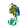
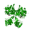
Mass: 65.409 Da / Num. of mol.: 6 / Source method: obtained synthetically / Formula: Zn
#3: Chemical ChemComp-BES / 2-(3-AMINO-2-HYDROXY-4-PHENYL-BUTYRYLAMINO)-4-METHYL-PENTANOIC ACID / BESTATIN / Ubenimex


Mass: 308.373 Da / Num. of mol.: 1 / Source method: obtained synthetically / Formula: C16H24N2O4 / Comment: protease inhibitor*YM
#4: Water ChemComp-HOH / water / Water


Mass: 18.015 Da / Num. of mol.: 590 / Source method: isolated from a natural source / Formula: H2O

-
Experimental details

-
Experiment

ExperimentMethod: X-RAY DIFFRACTION / Number of used crystals: 1

-
Sample preparation

CrystalDensity Matthews: 2.7 Å3/Da / Density % sol: 54.38 %
Crystal growTemperature: 293 K / Method: liquid diffusion / pH: 5.8
Details: 13% PEG 6000, 0.2M Zinc acetate, 0.1M MES-NaOH, pH 5.8

-
Data collection

DiffractionMean temperature: 90 K
Diffraction sourceSource: SYNCHROTRON / Site: SPring-8 / Beamline: BL44XU / Wavelength: 0.9 Å
DetectorType: BRUKER SMART 6000 / Detector: CCD / Date: May 16, 2012
RadiationProtocol: SINGLE WAVELENGTH / Monochromatic (M) / Laue (L): M / Scattering type: x-ray
Radiation wavelengthWavelength: 0.9 Å / Relative weight: 1
ReflectionResolution: 1.4→40 Å / Num. obs: 89717 / % possible obs: 93.4 % / Redundancy: 4.8 % / Net I/σ(I): 27.6

-
Processing

Software
NameVersionClassification
REFMAC5.5.0109refinement
MOSFLMdata reduction
SCALAdata scaling
MOLREPphasing
RefinementMethod to determine structure: MOLECULAR REPLACEMENT
Starting model: 2EK9
Resolution: 1.4→39.8 Å / Cor.coef. Fo:Fc: 0.966 / Cor.coef. Fo:Fc free: 0.944 / SU B: 1.195 / SU ML: 0.046 / Cross valid method: THROUGHOUT / ESU R: 0.06 / ESU R Free: 0.068 / Details: HYDROGENS HAVE BEEN ADDED IN THE RIDING POSITIONS
RfactorNum. reflection% reflectionSelection details
Rfree0.22284 4444 5 %RANDOM
Rwork0.1787 ---
obs0.18085 85042 92.93 %-
Solvent computationIon probe radii: 0.8 Å / Shrinkage radii: 0.8 Å / VDW probe radii: 1.4 Å
Displacement parametersBiso mean: 25.748 Å2
Baniso -1Baniso -2Baniso -3
1-0 Å2-0 Å2-0 Å2
2--0 Å20 Å2
3----0 Å2
Refinement stepCycle: 1 / Resolution: 1.4→39.8 Å
ProteinNucleic acidLigandSolventTotal
Num. atoms3119 0 28 590 3737
Refine LS restraints
Refine-IDTypeDev idealDev ideal targetNumber
X-RAY DIFFRACTIONr_bond_refined_d0.0260.0223192
X-RAY DIFFRACTIONr_bond_other_d
X-RAY DIFFRACTIONr_angle_refined_deg2.2681.9684319
X-RAY DIFFRACTIONr_angle_other_deg
X-RAY DIFFRACTIONr_dihedral_angle_1_deg6.2245411
X-RAY DIFFRACTIONr_dihedral_angle_2_deg36.59425.217138
X-RAY DIFFRACTIONr_dihedral_angle_3_deg14.8915545
X-RAY DIFFRACTIONr_dihedral_angle_4_deg16.0621515
X-RAY DIFFRACTIONr_chiral_restr0.1530.2493
X-RAY DIFFRACTIONr_gen_planes_refined0.0120.0212403
X-RAY DIFFRACTIONr_gen_planes_other
X-RAY DIFFRACTIONr_nbd_refined
X-RAY DIFFRACTIONr_nbd_other
X-RAY DIFFRACTIONr_nbtor_refined
X-RAY DIFFRACTIONr_nbtor_other
X-RAY DIFFRACTIONr_xyhbond_nbd_refined
X-RAY DIFFRACTIONr_xyhbond_nbd_other
X-RAY DIFFRACTIONr_metal_ion_refined
X-RAY DIFFRACTIONr_metal_ion_other
X-RAY DIFFRACTIONr_symmetry_vdw_refined
X-RAY DIFFRACTIONr_symmetry_vdw_other
X-RAY DIFFRACTIONr_symmetry_hbond_refined
X-RAY DIFFRACTIONr_symmetry_hbond_other
X-RAY DIFFRACTIONr_symmetry_metal_ion_refined
X-RAY DIFFRACTIONr_symmetry_metal_ion_other
X-RAY DIFFRACTIONr_mcbond_it1.4731.52038
X-RAY DIFFRACTIONr_mcbond_other
X-RAY DIFFRACTIONr_mcangle_it2.35323267
X-RAY DIFFRACTIONr_mcangle_other
X-RAY DIFFRACTIONr_scbond_it3.56831154
X-RAY DIFFRACTIONr_scbond_other
X-RAY DIFFRACTIONr_scangle_it5.9194.51052
X-RAY DIFFRACTIONr_scangle_other
X-RAY DIFFRACTIONr_long_range_B_refined
X-RAY DIFFRACTIONr_long_range_B_other
X-RAY DIFFRACTIONr_rigid_bond_restr
X-RAY DIFFRACTIONr_sphericity_free
X-RAY DIFFRACTIONr_sphericity_bonded
LS refinement shellResolution: 1.403→1.44 Å / Total num. of bins used: 20
RfactorNum. reflection% reflection
Rfree0.401 286 -
Rwork0.356 5334 -
obs--79.97 %

+
About Yorodumi

-
News

-
Feb 9, 2022. New format data for meta-information of EMDB entries

New format data for meta-information of EMDB entries

  • Version 3 of the EMDB header file is now the official format.
  • The previous official version 1.9 will be removed from the archive.

Related info.:EMDB header

External links:wwPDB to switch to version 3 of the EMDB data model

-
Aug 12, 2020. Covid-19 info

Covid-19 info

URL: https://pdbj.org/emnavi/covid19.php

New page: Covid-19 featured information page in EM Navigator.

Related info.:Covid-19 info / Mar 5, 2020. Novel coronavirus structure data

+
Mar 5, 2020. Novel coronavirus structure data

Novel coronavirus structure data

Related info.:Yorodumi Speices / Aug 12, 2020. Covid-19 info

External links:COVID-19 featured content - PDBj / Molecule of the Month (242):Coronavirus Proteases

+
Jan 31, 2019. EMDB accession codes are about to change! (news from PDBe EMDB page)

EMDB accession codes are about to change! (news from PDBe EMDB page)

  • The allocation of 4 digits for EMDB accession codes will soon come to an end. Whilst these codes will remain in use, new EMDB accession codes will include an additional digit and will expand incrementally as the available range of codes is exhausted. The current 4-digit format prefixed with “EMD-” (i.e. EMD-XXXX) will advance to a 5-digit format (i.e. EMD-XXXXX), and so on. It is currently estimated that the 4-digit codes will be depleted around Spring 2019, at which point the 5-digit format will come into force.
  • The EM Navigator/Yorodumi systems omit the EMD- prefix.

Related info.:Q: What is EMD? / ID/Accession-code notation in Yorodumi/EM Navigator

External links:EMDB Accession Codes are Changing Soon! / Contact to PDBj

+
Jul 12, 2017. Major update of PDB

Major update of PDB

  • wwPDB released updated PDB data conforming to the new PDBx/mmCIF dictionary.
  • This is a major update changing the version number from 4 to 5, and with Remediation, in which all the entries are updated.
  • In this update, many items about electron microscopy experimental information are reorganized (e.g. em_software).
  • Now, EM Navigator and Yorodumi are based on the updated data.

External links:wwPDB Remediation / Enriched Model Files Conforming to OneDep Data Standards Now Available in the PDB FTP Archive

-
Yorodumi

Thousand views of thousand structures

  • Yorodumi is a browser for structure data from EMDB, PDB, SASBDB, etc.
  • This page is also the successor to EM Navigator detail page, and also detail information page/front-end page for Omokage search.
  • The word "yorodu" (or yorozu) is an old Japanese word meaning "ten thousand". "mi" (miru) is to see.

Related info.:EMDB / PDB / SASBDB / Comparison of 3 databanks / Yorodumi Search / Aug 31, 2016. New EM Navigator & Yorodumi / Yorodumi Papers / Jmol/JSmol / Function and homology information / Changes in new EM Navigator and Yorodumi

Read more