 Movie
Movie Controller

[English] 日本語
 Yorodumi
- PDB-2cnb: Trypanosoma brucei UDP-galactose-4-epimerase in ternary complex w... -
+ Open data

- Basic information
| Entry | Database: PDB / ID: 2cnb | ||||||
|---|---|---|---|---|---|---|---|
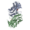
| Title | Trypanosoma brucei UDP-galactose-4-epimerase in ternary complex with NAD and the substrate analogue UDP-4-deoxy-4-fluoro-alpha-D-galactose | ||||||
 Components Components | UDP-GALACTOSE-4-EPIMERASE | ||||||
 Keywords Keywords |  EPIMERASE / SHORT-CHAIN DEHYDROGENASE/REDUCTASE / UDP-GALACTOSE-4- EPIMERASE / NAD / ISOMERASE / TRYPANOSOMA BRUCEI / UDP-4-DEOXY- 4-FLUORO-ALPHA-D-GALACTOSE EPIMERASE / SHORT-CHAIN DEHYDROGENASE/REDUCTASE / UDP-GALACTOSE-4- EPIMERASE / NAD / ISOMERASE / TRYPANOSOMA BRUCEI / UDP-4-DEOXY- 4-FLUORO-ALPHA-D-GALACTOSE | ||||||
| Function / homology |  Function and homology informationUDP-glucose 4-epimerase / UDP-glucose 4-epimerase activity / galactose metabolic process / glycosome / nucleotide binding Function and homology informationUDP-glucose 4-epimerase / UDP-glucose 4-epimerase activity / galactose metabolic process / glycosome / nucleotide bindingSimilarity search - Function | ||||||
| Biological species |  TRYPANOSOMA BRUCEI (eukaryote) TRYPANOSOMA BRUCEI (eukaryote) | ||||||
| Method | X-RAY DIFFRACTION / OTHER / Resolution: 2.7 Å | ||||||
 Authors Authors | Alphey, M.S. / Ferguson, M.A.J. / Hunter, W.N. | ||||||
 Citation Citation | Journal: Acta Crystallogr.,Sect.F / Year: 2006 Title: Trypanosoma Brucei Udp-Galactose-4-Epimerase in Ternary Complex with Nad+ and the Substrate Analogue Udp-4-Deoxy-4-Fluoro-Alpha-D-Galactose Authors: Alphey, M.S. / Burton, A. / Urbaniak, M. / Boons, G.J. / Ferguson, M.A.J. / Hunter, W.N. | ||||||
| History |
|
- Structure visualization
| Structure viewer | Molecule: MolmilJmol/JSmol |
|---|
- Downloads & links
-Download
| PDBx/mmCIF format | 2cnb.cif.gz | 306.3 KB | Display | PDBx/mmCIF format |
|---|---|---|---|---|
| PDB format | pdb2cnb.ent.gz | 249.5 KB | Display | PDB format |
| PDBx/mmJSON format | 2cnb.json.gz | Tree view | PDBx/mmJSON format | |
| Others |  Other downloads Other downloads |
-Validation report
| Arichive directory | https://data.pdbj.org/pub/pdb/validation_reports/cn/2cnbftp://data.pdbj.org/pub/pdb/validation_reports/cn/2cnb | HTTPS FTP |
|---|
-Related structure data
| Related structure data | |
|---|---|
| Similar structure data |


-Links
 PDBj
PDBj- Assembly
| Deposited unit | 
| ||||||||||||||||
|---|---|---|---|---|---|---|---|---|---|---|---|---|---|---|---|---|---|
| 1 | 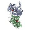
| ||||||||||||||||
| 2 | 
| ||||||||||||||||
| Unit cell |
| ||||||||||||||||
| Noncrystallographic symmetry (NCS) | NCS oper:
|
-Components
| #1: Protein | Mass: 43806.547 Da / Num. of mol.: 4 Source method: isolated from a genetically manipulated source Source: (gene. exp.) TRYPANOSOMA BRUCEI (eukaryote) / Plasmid: PET15B / Production host:  ESCHERICHIA COLI (E. coli) / Strain (production host): BL21 / References: UniProt: Q8T8E9, UDP-glucose 4-epimerase ESCHERICHIA COLI (E. coli) / Strain (production host): BL21 / References: UniProt: Q8T8E9, UDP-glucose 4-epimerase#2: Chemical | ChemComp-UFG /   Mass: 568.293 Da / Num. of mol.: 4 / Source method: obtained synthetically / Formula: C15H23FN2O16P2 Mass: 568.293 Da / Num. of mol.: 4 / Source method: obtained synthetically / Formula: C15H23FN2O16P2#3: Chemical | ChemComp-NAD / Nicotinamide adenine dinucleotide  Mass: 663.425 Da / Num. of mol.: 4 / Source method: obtained synthetically / Formula: C21H27N7O14P2 / Comment: NAD*YM Mass: 663.425 Da / Num. of mol.: 4 / Source method: obtained synthetically / Formula: C21H27N7O14P2 / Comment: NAD*YM#4: Water | ChemComp-HOH / | Water Mass: 18.015 Da / Num. of mol.: 416 / Source method: isolated from a natural source / Formula: H2O Mass: 18.015 Da / Num. of mol.: 416 / Source method: isolated from a natural source / Formula: H2OSequence details | SER -1 AND HIS 0 IN EACH SUBUNIT CORRESPOND TO A VISIBLE FRAGMENT OF THE 6HIS AFFINITY TAG USED FOR ...SER -1 AND HIS 0 IN EACH SUBUNIT CORRESPOND | |
|---|
-Experimental details
-Experiment
| Experiment | Method: X-RAY DIFFRACTION / Number of used crystals: 1 |
|---|
- Sample preparation
| Crystal | Density Matthews: 3 Å3/Da / Density % sol: 58.7 % |
|---|---|
| Crystal grow | pH: 8 Details: 8% PEG 8000, 200MM KCL, 50MM NA2B4O7, 5% GLYCEROL, PH 8.5 |
-Data collection
| Diffraction | Mean temperature: 100 K |
|---|---|
| Diffraction source | Source: ROTATING ANODE / Type: RIGAKU MICROMAX-007 HF / Wavelength: 1.5418 |
| Detector | Type: RIGAKU IMAGE PLATE / Detector: IMAGE PLATE / Date: Nov 11, 2005 / Details: MIRRORS |
| Radiation | Protocol: SINGLE WAVELENGTH / Monochromatic (M) / Laue (L): M / Scattering type: x-ray |
| Radiation wavelength | Wavelength: 1.5418 Å / Relative weight: 1 |
| Reflection | Resolution: 2.7→92 Å / Num. obs: 49501 / % possible obs: 96.8 % / Observed criterion σ(I): 2 / Redundancy: 4.5 % / Biso Wilson estimate: 64.7 Å2 / Rmerge(I) obs: 0.09 / Net I/σ(I): 15.2 |
| Reflection shell | Resolution: 2.7→2.77 Å / Redundancy: 3.3 % / Rmerge(I) obs: 0.47 / Mean I/σ(I) obs: 2.4 / % possible all: 78.9 |
- Processing
| Software |
| ||||||||||||||||||||||||||||||||||||||||||||||||||||||||||||||||||||||||||||||||||||||||||||||||||||||||||||||||||||||||||||||||||||||||||||||||||||||||||||||||||||||||||||||||||||||
|---|---|---|---|---|---|---|---|---|---|---|---|---|---|---|---|---|---|---|---|---|---|---|---|---|---|---|---|---|---|---|---|---|---|---|---|---|---|---|---|---|---|---|---|---|---|---|---|---|---|---|---|---|---|---|---|---|---|---|---|---|---|---|---|---|---|---|---|---|---|---|---|---|---|---|---|---|---|---|---|---|---|---|---|---|---|---|---|---|---|---|---|---|---|---|---|---|---|---|---|---|---|---|---|---|---|---|---|---|---|---|---|---|---|---|---|---|---|---|---|---|---|---|---|---|---|---|---|---|---|---|---|---|---|---|---|---|---|---|---|---|---|---|---|---|---|---|---|---|---|---|---|---|---|---|---|---|---|---|---|---|---|---|---|---|---|---|---|---|---|---|---|---|---|---|---|---|---|---|---|---|---|---|---|
| Refinement | Method to determine structure: OTHER / Resolution: 2.7→91.67 Å / Cor.coef. Fo:Fc: 0.954 / Cor.coef. Fo:Fc free: 0.906 / SU B: 15.525 / SU ML: 0.309 / Cross valid method: THROUGHOUT / σ(F): 0 / ESU R Free: 0.402 / Stereochemistry target values: MAXIMUM LIKELIHOOD / Details: HYDROGENS HAVE BEEN ADDED IN THE RIDING POSITIONS.
| ||||||||||||||||||||||||||||||||||||||||||||||||||||||||||||||||||||||||||||||||||||||||||||||||||||||||||||||||||||||||||||||||||||||||||||||||||||||||||||||||||||||||||||||||||||||
| Solvent computation | Ion probe radii: 0.8 Å / VDW probe radii: 1.2 Å / Solvent model: MASK BULK SOLVENT | ||||||||||||||||||||||||||||||||||||||||||||||||||||||||||||||||||||||||||||||||||||||||||||||||||||||||||||||||||||||||||||||||||||||||||||||||||||||||||||||||||||||||||||||||||||||
| Displacement parameters | Biso mean: 48.29 Å2
| ||||||||||||||||||||||||||||||||||||||||||||||||||||||||||||||||||||||||||||||||||||||||||||||||||||||||||||||||||||||||||||||||||||||||||||||||||||||||||||||||||||||||||||||||||||||
| Refinement step | Cycle: LAST / Resolution: 2.7→91.67 Å
| ||||||||||||||||||||||||||||||||||||||||||||||||||||||||||||||||||||||||||||||||||||||||||||||||||||||||||||||||||||||||||||||||||||||||||||||||||||||||||||||||||||||||||||||||||||||
| Refine LS restraints |
|