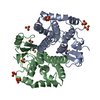
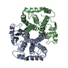
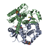
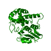
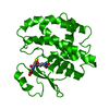
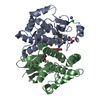
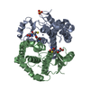
[English] 日本語
Yorodumi
- PDB-4l5o: Crystal structure of 26 kDa GST D26H mutant of Clonorchis sinensis -

+
Open data


ID or keywords:

Loading...

-
Basic information

Entry
Database: PDB / ID: 4l5o
TitleCrystal structure of 26 kDa GST D26H mutant of Clonorchis sinensis
ComponentsPutative glutathione transferaseGlutathione S-transferase
KeywordsTRANSFERASE / GSH binding
Function / homology
Function and homology information


glutathione transferase / glutathione transferase activity / glutathione metabolic process / metal ion binding
Similarity search - Function
Glutathione S-transferase, C-terminal domain / Glutathione S-transferase, N-terminal domain / Glutathione transferase family / Glutathione S-transferase Yfyf (Class Pi); Chain A, domain 2 - #10 / Glutathione S-transferase, C-terminal / Glutathione S-transferase Yfyf (Class Pi); Chain A, domain 2 / Soluble glutathione S-transferase N-terminal domain profile. / Glutathione S-transferase, C-terminal-like / Soluble glutathione S-transferase C-terminal domain profile. / Glutathione S-transferase, N-terminal ...Glutathione S-transferase, C-terminal domain / Glutathione S-transferase, N-terminal domain / Glutathione transferase family / Glutathione S-transferase Yfyf (Class Pi); Chain A, domain 2 - #10 / Glutathione S-transferase, C-terminal / Glutathione S-transferase Yfyf (Class Pi); Chain A, domain 2 / Soluble glutathione S-transferase N-terminal domain profile. / Glutathione S-transferase, C-terminal-like / Soluble glutathione S-transferase C-terminal domain profile. / Glutathione S-transferase, N-terminal / Glutathione S-transferase, C-terminal domain superfamily / Glutaredoxin / Glutaredoxin / Thioredoxin-like superfamily / Up-down Bundle / 3-Layer(aba) Sandwich / Mainly Alpha / Alpha Beta
Similarity search - Domain/homology
GLUTATHIONE / glutathione transferase
Similarity search - Component
Biological speciesClonorchis sinensis (oriental liver fluke)
MethodX-RAY DIFFRACTION / SYNCHROTRON / MOLECULAR REPLACEMENT / molecular replacement / Resolution: 2.09 Å
AuthorsChung, Y.J. / Han, Y.H.
CitationJournal: Biochem.Biophys.Res.Commun. / Year: 2013
Title: Crystal structures of 26kDa Clonorchis sinensis glutathione S-transferase reveal zinc binding and putative metal binding.
Authors: Han, Y.H. / Hong, S.J. / Cheong, H.K. / Chung, Y.J.
History
DepositionJun 11, 2013Deposition site: RCSB / Processing site: PDBJ
Revision 1.0Sep 11, 2013Provider: repository / Type: Initial release
Revision 1.1Jun 17, 2015Group: Database references
Revision 1.2Nov 15, 2017Group: Refinement description / Category: software
Revision 1.3Mar 20, 2024Group: Data collection / Database references / Derived calculations
Category: chem_comp_atom / chem_comp_bond ...chem_comp_atom / chem_comp_bond / database_2 / pdbx_struct_conn_angle / struct_conn / struct_ref_seq_dif / struct_site
Item: _database_2.pdbx_DOI / _database_2.pdbx_database_accession ..._database_2.pdbx_DOI / _database_2.pdbx_database_accession / _pdbx_struct_conn_angle.ptnr1_auth_asym_id / _pdbx_struct_conn_angle.ptnr1_auth_seq_id / _pdbx_struct_conn_angle.ptnr1_label_asym_id / _pdbx_struct_conn_angle.ptnr1_label_seq_id / _pdbx_struct_conn_angle.ptnr3_auth_asym_id / _pdbx_struct_conn_angle.ptnr3_auth_seq_id / _pdbx_struct_conn_angle.ptnr3_label_asym_id / _pdbx_struct_conn_angle.ptnr3_label_seq_id / _pdbx_struct_conn_angle.value / _struct_conn.pdbx_dist_value / _struct_conn.ptnr1_auth_asym_id / _struct_conn.ptnr1_auth_comp_id / _struct_conn.ptnr1_auth_seq_id / _struct_conn.ptnr1_label_asym_id / _struct_conn.ptnr1_label_atom_id / _struct_conn.ptnr1_label_comp_id / _struct_conn.ptnr1_label_seq_id / _struct_conn.ptnr2_auth_asym_id / _struct_conn.ptnr2_auth_comp_id / _struct_conn.ptnr2_auth_seq_id / _struct_conn.ptnr2_label_asym_id / _struct_conn.ptnr2_label_atom_id / _struct_conn.ptnr2_label_comp_id / _struct_conn.ptnr2_label_seq_id / _struct_ref_seq_dif.details / _struct_site.pdbx_auth_asym_id / _struct_site.pdbx_auth_comp_id / _struct_site.pdbx_auth_seq_id

-
Structure visualization

Structure viewerMolecule:
MolmilJmol/JSmol

Downloads & links

-
Assembly

Deposited unit
A: Putative glutathione transferase
B: Putative glutathione transferase
C: Putative glutathione transferase
hetero molecules


Theoretical massNumber of molelcules
Total (without water)76,89514
Polymers75,2663
Non-polymers1,62911
Water4,594255
1
A: Putative glutathione transferase
B: Putative glutathione transferase
hetero molecules


Theoretical massNumber of molelcules
Total (without water)51,1158
Polymers50,1772
Non-polymers9386
Water362
TypeNameSymmetry operationNumber
identity operation1_555x,y,z1
Buried area4040 Å2
ΔGint-44 kcal/mol
Surface area19480 Å2
MethodPISA
2
C: Putative glutathione transferase
hetero molecules

C: Putative glutathione transferase
hetero molecules


Theoretical massNumber of molelcules
Total (without water)51,56012
Polymers50,1772
Non-polymers1,38310
Water362
TypeNameSymmetry operationNumber
identity operation1_555x,y,z1
crystal symmetry operation6_555-x,-x+y,-z+1/31
Buried area4820 Å2
ΔGint-98 kcal/mol
Surface area20230 Å2
MethodPISA
Unit cell
Length a, b, c (Å)98.370, 98.370, 178.501
Angle α, β, γ (deg.)90.00, 90.00, 120.00
Int Tables number152
Space group name H-MP3121
DetailsThe one biological dimer and one monomer are in asymmetric unit. The biological assembly of the monomer is generated by the three fold axis : -x, -x+y, -z+1/3

-
Components

#1: Protein Putative glutathione transferase / Glutathione S-transferase


Mass: 25088.648 Da / Num. of mol.: 3 / Mutation: D26H
Source method: isolated from a genetically manipulated source
Source: (gene. exp.) Clonorchis sinensis (oriental liver fluke)
Plasmid: pET25b / Production host: Escherichia coli (E. coli) / Strain (production host): BL21(DE3) / References: UniProt: Q25595
#2: Chemical ChemComp-GSH / GLUTATHIONE / Glutathione


Mass: 307.323 Da / Num. of mol.: 3 / Source method: obtained synthetically / Formula: C10H17N3O6S
#3: Chemical
ChemComp-SO4 / SULFATE ION / Sulfate


Mass: 96.063 Da / Num. of mol.: 6 / Source method: obtained synthetically / Formula: SO4
#4: Chemical ChemComp-ZN / ZINC ION


Mass: 65.409 Da / Num. of mol.: 2 / Source method: obtained synthetically / Formula: Zn
#5: Water ChemComp-HOH / water / Water


Mass: 18.015 Da / Num. of mol.: 255 / Source method: isolated from a natural source / Formula: H2O

-
Experimental details

-
Experiment

ExperimentMethod: X-RAY DIFFRACTION / Number of used crystals: 1

-
Sample preparation

CrystalDensity Matthews: 3.31 Å3/Da / Density % sol: 62.87 %
Crystal growTemperature: 293 K / Method: vapor diffusion, hanging drop / pH: 8.5
Details: 0.1M Tris pH8.5, 5mM Zinc sulfate, 2.0M Ammonium sulfate, VAPOR DIFFUSION, HANGING DROP, temperature 293K

-
Data collection

DiffractionMean temperature: 100 K
Diffraction sourceSource: SYNCHROTRON / Site: PAL/PLS / Beamline: 4A / Wavelength: 1 Å
DetectorType: ADSC QUANTUM 315r / Detector: CCD / Date: Jul 16, 2009
RadiationProtocol: SINGLE WAVELENGTH / Monochromatic (M) / Laue (L): M / Scattering type: x-ray
Radiation wavelengthWavelength: 1 Å / Relative weight: 1
ReflectionResolution: 2.089→85.191 Å / Num. all: 60028 / Num. obs: 60028 / % possible obs: 100 % / Redundancy: 5.9 % / Rsym value: 0.193 / Net I/σ(I): 7.2
Reflection shell
Resolution (Å)Redundancy (%)Rmerge(I) obsMean I/σ(I) obsRsym valueDiffraction-ID% possible all
2.09-2.25.70.0190.41.928199.9
2.2-2.345.80.0190.61.2931100
2.34-2.55.90.0190.90.891100
2.5-2.760.0191.20.6231100
2.7-2.9560.01920.3781100
2.95-3.360.0193.70.2031100
3.3-3.815.90.0197.10.1031100
3.81-4.675.80.0198.10.0781100
4.67-6.615.50.0199.70.063199.9
6.61-59.55.70.01919.80.029199.8

-
Phasing

PhasingMethod: molecular replacement
Phasing MR
Highest resolutionLowest resolution
Rotation2.7 Å59.5 Å
Translation2.7 Å59.5 Å

-
Processing

Software
NameVersionClassificationNB
SCALA3.3.9data scaling
MOLREPphasing
REFMACrefinement
PDB_EXTRACT3.11data extraction
HKL-2000data collection
DENZOdata reduction
SCALEPACKdata scaling
RefinementMethod to determine structure: MOLECULAR REPLACEMENT / Resolution: 2.09→59.57 Å / Cor.coef. Fo:Fc: 0.955 / Cor.coef. Fo:Fc free: 0.935 / Occupancy max: 1 / Occupancy min: 0.4 / SU B: 5.623 / SU ML: 0.141 / SU R Cruickshank DPI: 0.1738 / Cross valid method: THROUGHOUT / ESU R: 0.174 / ESU R Free: 0.166 / Stereochemistry target values: MAXIMUM LIKELIHOOD / Details: HYDROGENS HAVE BEEN ADDED IN THE RIDING POSITIONS
RfactorNum. reflection% reflectionSelection details
Rfree0.24818 3029 5.1 %RANDOM
Rwork0.20171 ---
obs0.20409 56931 99.94 %-
Solvent computationIon probe radii: 0.8 Å / Shrinkage radii: 0.8 Å / VDW probe radii: 1.2 Å / Solvent model: MASK
Displacement parametersBiso mean: 39.257 Å2
Baniso -1Baniso -2Baniso -3
1-0.26 Å20.26 Å20 Å2
2--0.26 Å20 Å2
3----0.85 Å2
Refinement stepCycle: LAST / Resolution: 2.09→59.57 Å
ProteinNucleic acidLigandSolventTotal
Num. atoms5286 0 92 255 5633
Refine LS restraints
Refine-IDTypeDev idealDev ideal targetNumber
X-RAY DIFFRACTIONr_bond_refined_d0.0160.0195538
X-RAY DIFFRACTIONr_bond_other_d0.0010.025129
X-RAY DIFFRACTIONr_angle_refined_deg1.7681.9667518
X-RAY DIFFRACTIONr_angle_other_deg0.8433.00211752
X-RAY DIFFRACTIONr_dihedral_angle_1_deg6.0115650
X-RAY DIFFRACTIONr_dihedral_angle_2_deg33.18423.582268
X-RAY DIFFRACTIONr_dihedral_angle_3_deg15.79315892
X-RAY DIFFRACTIONr_dihedral_angle_4_deg17.6711533
X-RAY DIFFRACTIONr_chiral_restr0.1010.2775
X-RAY DIFFRACTIONr_gen_planes_refined0.0090.0216251
X-RAY DIFFRACTIONr_gen_planes_other0.0010.021344
X-RAY DIFFRACTIONr_nbd_refined
X-RAY DIFFRACTIONr_nbd_other
X-RAY DIFFRACTIONr_nbtor_refined
X-RAY DIFFRACTIONr_nbtor_other
X-RAY DIFFRACTIONr_xyhbond_nbd_refined
X-RAY DIFFRACTIONr_xyhbond_nbd_other
X-RAY DIFFRACTIONr_metal_ion_refined
X-RAY DIFFRACTIONr_metal_ion_other
X-RAY DIFFRACTIONr_symmetry_vdw_refined
X-RAY DIFFRACTIONr_symmetry_vdw_other
X-RAY DIFFRACTIONr_symmetry_hbond_refined
X-RAY DIFFRACTIONr_symmetry_hbond_other
X-RAY DIFFRACTIONr_symmetry_metal_ion_refined
X-RAY DIFFRACTIONr_symmetry_metal_ion_other
X-RAY DIFFRACTIONr_mcbond_it
X-RAY DIFFRACTIONr_mcbond_other
X-RAY DIFFRACTIONr_mcangle_it
X-RAY DIFFRACTIONr_mcangle_other
X-RAY DIFFRACTIONr_scbond_it
X-RAY DIFFRACTIONr_scbond_other
X-RAY DIFFRACTIONr_scangle_it
X-RAY DIFFRACTIONr_scangle_other
X-RAY DIFFRACTIONr_long_range_B_refined
X-RAY DIFFRACTIONr_long_range_B_other
X-RAY DIFFRACTIONr_rigid_bond_restr
X-RAY DIFFRACTIONr_sphericity_free
X-RAY DIFFRACTIONr_sphericity_bonded
LS refinement shellResolution: 2.089→2.143 Å / Total num. of bins used: 20
RfactorNum. reflection% reflection
Rfree0.375 236 -
Rwork0.342 4125 -
obs--99.54 %

+
About Yorodumi

-
News

-
Feb 9, 2022. New format data for meta-information of EMDB entries

New format data for meta-information of EMDB entries

  • Version 3 of the EMDB header file is now the official format.
  • The previous official version 1.9 will be removed from the archive.

Related info.:EMDB header

External links:wwPDB to switch to version 3 of the EMDB data model

-
Aug 12, 2020. Covid-19 info

Covid-19 info

URL: https://pdbj.org/emnavi/covid19.php

New page: Covid-19 featured information page in EM Navigator.

Related info.:Covid-19 info / Mar 5, 2020. Novel coronavirus structure data

+
Mar 5, 2020. Novel coronavirus structure data

Novel coronavirus structure data

Related info.:Yorodumi Speices / Aug 12, 2020. Covid-19 info

External links:COVID-19 featured content - PDBj / Molecule of the Month (242):Coronavirus Proteases

+
Jan 31, 2019. EMDB accession codes are about to change! (news from PDBe EMDB page)

EMDB accession codes are about to change! (news from PDBe EMDB page)

  • The allocation of 4 digits for EMDB accession codes will soon come to an end. Whilst these codes will remain in use, new EMDB accession codes will include an additional digit and will expand incrementally as the available range of codes is exhausted. The current 4-digit format prefixed with “EMD-” (i.e. EMD-XXXX) will advance to a 5-digit format (i.e. EMD-XXXXX), and so on. It is currently estimated that the 4-digit codes will be depleted around Spring 2019, at which point the 5-digit format will come into force.
  • The EM Navigator/Yorodumi systems omit the EMD- prefix.

Related info.:Q: What is EMD? / ID/Accession-code notation in Yorodumi/EM Navigator

External links:EMDB Accession Codes are Changing Soon! / Contact to PDBj

+
Jul 12, 2017. Major update of PDB

Major update of PDB

  • wwPDB released updated PDB data conforming to the new PDBx/mmCIF dictionary.
  • This is a major update changing the version number from 4 to 5, and with Remediation, in which all the entries are updated.
  • In this update, many items about electron microscopy experimental information are reorganized (e.g. em_software).
  • Now, EM Navigator and Yorodumi are based on the updated data.

External links:wwPDB Remediation / Enriched Model Files Conforming to OneDep Data Standards Now Available in the PDB FTP Archive

-
Yorodumi

Thousand views of thousand structures

  • Yorodumi is a browser for structure data from EMDB, PDB, SASBDB, etc.
  • This page is also the successor to EM Navigator detail page, and also detail information page/front-end page for Omokage search.
  • The word "yorodu" (or yorozu) is an old Japanese word meaning "ten thousand". "mi" (miru) is to see.

Related info.:EMDB / PDB / SASBDB / Comparison of 3 databanks / Yorodumi Search / Aug 31, 2016. New EM Navigator & Yorodumi / Yorodumi Papers / Jmol/JSmol / Function and homology information / Changes in new EM Navigator and Yorodumi

Read more