 Movie
Movie Controller

[English] 日本語
 Yorodumi
- PDB-2boj: crystal Structure of pseudomonas aeruginosa lectin (PA-IIL) compl... -
+ Open data

- Basic information
| Entry | Database: PDB / ID: 2boj | ||||||
|---|---|---|---|---|---|---|---|
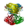
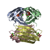
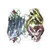
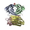
| Title | crystal Structure of pseudomonas aeruginosa lectin (PA-IIL) complexed with methyl-B-D-Arabinopyranoside | ||||||
 Components Components | PSEUDOMONAS AERUGINOSA LECTIN II | ||||||
 Keywords Keywords |  LECTIN / ARABINOSE / CALCIUM / LEWIS A / CYSTIC FIBROSIS LECTIN / ARABINOSE / CALCIUM / LEWIS A / CYSTIC FIBROSIS | ||||||
| Function / homology |  Function and homology information Function and homology informationsingle-species biofilm formation / carbohydrate binding / metal ion bindingSimilarity search - Function | ||||||
| Biological species |   PSEUDOMONAS AERUGINOSA (bacteria) PSEUDOMONAS AERUGINOSA (bacteria) | ||||||
| Method | X-RAY DIFFRACTION / SYNCHROTRON / MOLECULAR REPLACEMENT / Resolution: 1.8 Å | ||||||
 Authors Authors | Sabin, C.D. / Mitchell, E.P. / Wimmerova, M. / Imberty, A. | ||||||
 Citation Citation | Journal: FEBS Lett. / Year: 2006 Title: Binding of Different Monosaccharides by Lectin Pa-Iil from Pseudoman Aeruginosa: Thermodynamics Data Correlated with X-Ray Structures. Authors: Sabin, C.D. / Mitchell, E.P. / Pokarna, M. / Gautier, C. / Utille, J.-P. / Wimmerova, M. / Imberty, A. #1: Journal: Proteins: Struct.,Funct., Genet. / Year: 2005Title: High Affinity Fucose Binding of Pseudomonas Aeruginosa Lectin Pa-Iil: 1.0 A Resolution Crystal Structure of the Complex Combined with Thermodynamics and Computational Chemistry Approaches Authors: Mitchell, E. / Sabin, C.D. / Snajdrova, L. / Pokorna, M. / Perret, S. / Gautier, C. / Hofr, C. / Gilboa-Garber, N. / Koca, J. / Wimmerova, M. / Imberty, A. | ||||||
| History |
| ||||||
| Remark 700 | SHEET DETERMINATION METHOD: AUTHOR PROVIDED. |
- Structure visualization
| Structure viewer | Molecule: MolmilJmol/JSmol |
|---|
- Downloads & links
-Download
| PDBx/mmCIF format | 2boj.cif.gz | 114.2 KB | Display | PDBx/mmCIF format |
|---|---|---|---|---|
| PDB format | pdb2boj.ent.gz | 87.8 KB | Display | PDB format |
| PDBx/mmJSON format | 2boj.json.gz | Tree view | PDBx/mmJSON format | |
| Others |  Other downloads Other downloads |
-Validation report
| Arichive directory | https://data.pdbj.org/pub/pdb/validation_reports/bo/2bojftp://data.pdbj.org/pub/pdb/validation_reports/bo/2boj | HTTPS FTP |
|---|
-Related structure data
| Related structure data |  2bp6C  1uzvS  1w43 S: Starting model for refinement C: citing same article ( |
|---|---|
| Similar structure data |


-Links
 PDBj
PDBj- Assembly
| Deposited unit | 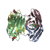
| ||||||||
|---|---|---|---|---|---|---|---|---|---|
| 1 |
| ||||||||
| Unit cell |
|
-Components
| #1: Protein | Mass: 11734.707 Da / Num. of mol.: 4 Source method: isolated from a genetically manipulated source Source: (gene. exp.) PSEUDOMONAS AERUGINOSA (bacteria) / Strain: PAO1 / Plasmid: PET25PAIIL / Production host: ESCHERICHIA COLI (E. coli) / Strain (production host): BL21(DE3) / References: UniProt: Q9HYN5#2: Sugar | ChemComp-ARW /   Type: D-saccharide / Mass: 164.156 Da / Num. of mol.: 4 / Source method: obtained synthetically / Formula: C6H12O5 Type: D-saccharide / Mass: 164.156 Da / Num. of mol.: 4 / Source method: obtained synthetically / Formula: C6H12O5#3: Chemical | ChemComp-CA /   Mass: 40.078 Da / Num. of mol.: 8 / Source method: obtained synthetically / Formula: Ca Mass: 40.078 Da / Num. of mol.: 8 / Source method: obtained synthetically / Formula: Ca#4: Chemical | ChemComp-SO4 / | Sulfate  Mass: 96.063 Da / Num. of mol.: 1 / Source method: obtained synthetically / Formula: SO4 Mass: 96.063 Da / Num. of mol.: 1 / Source method: obtained synthetically / Formula: SO4#5: Water | ChemComp-HOH / | Water Mass: 18.015 Da / Num. of mol.: 612 / Source method: isolated from a natural source / Formula: H2O Mass: 18.015 Da / Num. of mol.: 612 / Source method: isolated from a natural source / Formula: H2O |
|---|
-Experimental details
-Experiment
| Experiment | Method: X-RAY DIFFRACTION / Number of used crystals: 1 |
|---|
- Sample preparation
| Crystal | Density Matthews: 1.74 Å3/Da / Density % sol: 29 % |
|---|---|
| Crystal grow | pH: 8.5 Details: TRIS HCL 0.1M, PH8.5, 1.75 M AMMONIUM SULFATE, pH 8.50 |
-Data collection
| Diffraction | Mean temperature: 100 K |
|---|---|
| Diffraction source | Source: SYNCHROTRON / Site: ESRF  / Beamline: ID14-2 / Wavelength: 0.933 / Beamline: ID14-2 / Wavelength: 0.933 |
| Detector | Type: ADSC CCD / Detector: CCD / Date: Jun 10, 2002 / Details: TOROIDAL MIRROR |
| Radiation | Monochromator: SINGLE CRYSTAL DIAMOND / Protocol: SINGLE WAVELENGTH / Monochromatic (M) / Laue (L): M / Scattering type: x-ray |
| Radiation wavelength | Wavelength: 0.933 Å / Relative weight: 1 |
| Reflection | Resolution: 1.8→29.6 Å / Num. obs: 35769 / % possible obs: 97.9 % / Redundancy: 3.73 % / Rmerge(I) obs: 0.07 / Net I/σ(I): 7.5 |
| Reflection shell | Resolution: 1.8→1.9 Å / Redundancy: 3.45 % / Rmerge(I) obs: 0.19 / Mean I/σ(I) obs: 3.64 / % possible all: 89 |
- Processing
| Software |
| ||||||||||||||||||||||||||||||||||||||||||||||||||||||||||||||||||||||||||||||||||||||||||||||||||||||||||||||||||||||||||||||||||||||||||||||||||||||||||||||||||||||||||||||||||||||
|---|---|---|---|---|---|---|---|---|---|---|---|---|---|---|---|---|---|---|---|---|---|---|---|---|---|---|---|---|---|---|---|---|---|---|---|---|---|---|---|---|---|---|---|---|---|---|---|---|---|---|---|---|---|---|---|---|---|---|---|---|---|---|---|---|---|---|---|---|---|---|---|---|---|---|---|---|---|---|---|---|---|---|---|---|---|---|---|---|---|---|---|---|---|---|---|---|---|---|---|---|---|---|---|---|---|---|---|---|---|---|---|---|---|---|---|---|---|---|---|---|---|---|---|---|---|---|---|---|---|---|---|---|---|---|---|---|---|---|---|---|---|---|---|---|---|---|---|---|---|---|---|---|---|---|---|---|---|---|---|---|---|---|---|---|---|---|---|---|---|---|---|---|---|---|---|---|---|---|---|---|---|---|---|
| Refinement | Method to determine structure: MOLECULAR REPLACEMENT Starting model: PDB ENTRY 1UZV Resolution: 1.8→49.39 Å / Cor.coef. Fo:Fc: 0.964 / Cor.coef. Fo:Fc free: 0.945 / SU B: 2.129 / SU ML: 0.068 / Cross valid method: THROUGHOUT / ESU R: 0.118 / ESU R Free: 0.108 / Stereochemistry target values: MAXIMUM LIKELIHOOD Details: HYDROGENS HAVE BEEN ADDED IN THE RIDING POSITIONS. DISORDERED SIDE CHAINS ATOMS WERE MODELED WITH OCCUPANCIES LESS THAN 1.0
| ||||||||||||||||||||||||||||||||||||||||||||||||||||||||||||||||||||||||||||||||||||||||||||||||||||||||||||||||||||||||||||||||||||||||||||||||||||||||||||||||||||||||||||||||||||||
| Solvent computation | Ion probe radii: 0.8 Å / Shrinkage radii: 0.8 Å / VDW probe radii: 1.2 Å / Solvent model: MASK | ||||||||||||||||||||||||||||||||||||||||||||||||||||||||||||||||||||||||||||||||||||||||||||||||||||||||||||||||||||||||||||||||||||||||||||||||||||||||||||||||||||||||||||||||||||||
| Displacement parameters | Biso mean: 11.43 Å2
| ||||||||||||||||||||||||||||||||||||||||||||||||||||||||||||||||||||||||||||||||||||||||||||||||||||||||||||||||||||||||||||||||||||||||||||||||||||||||||||||||||||||||||||||||||||||
| Refinement step | Cycle: LAST / Resolution: 1.8→49.39 Å
| ||||||||||||||||||||||||||||||||||||||||||||||||||||||||||||||||||||||||||||||||||||||||||||||||||||||||||||||||||||||||||||||||||||||||||||||||||||||||||||||||||||||||||||||||||||||
| Refine LS restraints |
|