 Movie
Movie Controller


+ Open data

- Basic information
| Entry | Database: PDB / ID: 1ovs | |||||||||
|---|---|---|---|---|---|---|---|---|---|---|
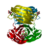
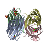
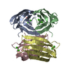
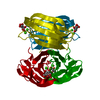
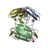
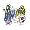
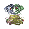
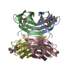
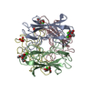
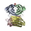
| Title | LecB (PA-LII) in complex with core trimannoside | |||||||||
 Components Components | hypothetical protein LecB Hypothesis Hypothesis | |||||||||
 Keywords Keywords | SUGAR BINDING PROTEIN / lectin / carbohydrate | |||||||||
| Function / homology |  Function and homology information Function and homology informationsingle-species biofilm formation / carbohydrate binding / metal ion bindingSimilarity search - Function | |||||||||
| Biological species |   Pseudomonas aeruginosa (bacteria) Pseudomonas aeruginosa (bacteria) | |||||||||
| Method | X-RAY DIFFRACTION / SYNCHROTRON / MOLECULAR REPLACEMENT / Resolution: 1.75 Å | |||||||||
 Authors Authors | Loris, R. / Tielker, D. / Jaeger, K.-E. / Wyns, L. | |||||||||
 Citation Citation | Journal: J.MOL.BIOL. / Year: 2003 Title: Structural Basis of Carbohydrate Recognition by the Lectin LecB from Pseudomonas aeruginosa Authors: Loris, R. / Tielker, D. / Jaeger, K.-E. / Wyns, L. | |||||||||
| History |
|
- Structure visualization
| Structure viewer | Molecule: MolmilJmol/JSmol |
|---|
- Downloads & links
-Download
| PDBx/mmCIF format | 1ovs.cif.gz | 101.4 KB | Display | PDBx/mmCIF format |
|---|---|---|---|---|
| PDB format | pdb1ovs.ent.gz | 78.1 KB | Display | PDB format |
| PDBx/mmJSON format | 1ovs.json.gz | Tree view | PDBx/mmJSON format | |
| Others |  Other downloads Other downloads |
-Validation report
| Arichive directory | https://data.pdbj.org/pub/pdb/validation_reports/ov/1ovsftp://data.pdbj.org/pub/pdb/validation_reports/ov/1ovs | HTTPS FTP |
|---|
-Related structure data


-Links
 PDBj
PDBj


- Assembly
| Deposited unit | 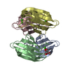
| ||||||||
|---|---|---|---|---|---|---|---|---|---|
| 1 |
| ||||||||
| 2 | 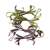
| ||||||||
| 3 | 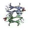
| ||||||||
| Unit cell |
|
-Components
| #1: Protein | Hypothesis / LecB Mass: 11734.707 Da / Num. of mol.: 4 Source method: isolated from a genetically manipulated source Source: (gene. exp.) Pseudomonas aeruginosa (bacteria) / Gene: LecB / Plasmid: pET22a / Production host: Escherichia coli (E. coli) / References: GenBank: 9949495, UniProt: Q9HYN5*PLUS#2: Polysaccharide | 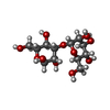  , Oligosaccharide / Class: Metabolism / Mass: 342.297 Da / Num. of mol.: 2 , Oligosaccharide / Class: Metabolism / Mass: 342.297 Da / Num. of mol.: 2Source method: isolated from a genetically manipulated source Details: oligosaccharide / References: 3alpha-alpha-mannobiose #3: Chemical | ChemComp-CA /   Mass: 40.078 Da / Num. of mol.: 8 / Source method: obtained synthetically / Formula: Ca Mass: 40.078 Da / Num. of mol.: 8 / Source method: obtained synthetically / Formula: Ca#4: Sugar | Mannose  Type: D-saccharide, alpha linking / Mass: 180.156 Da / Num. of mol.: 2 Type: D-saccharide, alpha linking / Mass: 180.156 Da / Num. of mol.: 2Source method: isolated from a genetically manipulated source Formula: C6H12O6 #5: Water | ChemComp-HOH / | Water Mass: 18.015 Da / Num. of mol.: 324 / Source method: isolated from a natural source / Formula: H2O Mass: 18.015 Da / Num. of mol.: 324 / Source method: isolated from a natural source / Formula: H2O |
|---|
-Experimental details
-Experiment
| Experiment | Method: X-RAY DIFFRACTION / Number of used crystals: 1 |
|---|
- Sample preparation
| Crystal | Density Matthews: 1.73 Å3/Da / Density % sol: 28.26 % | ||||||||||||||||||||||||||||||
|---|---|---|---|---|---|---|---|---|---|---|---|---|---|---|---|---|---|---|---|---|---|---|---|---|---|---|---|---|---|---|---|
| Crystal grow | Temperature: 293 K / Method: vapor diffusion, hanging drop / pH: 8 Details: PEG8000, ammonium sulphate, pH 8.0, VAPOR DIFFUSION, HANGING DROP, temperature 293K | ||||||||||||||||||||||||||||||
| Crystal grow | *PLUS Method: vapor diffusion, hanging drop | ||||||||||||||||||||||||||||||
| Components of the solutions | *PLUS
|
-Data collection
| Diffraction | Mean temperature: 293 K |
|---|---|
| Diffraction source | Source: SYNCHROTRON / Site: ESRF  / Beamline: ID14-2 / Wavelength: 0.933 Å / Beamline: ID14-2 / Wavelength: 0.933 Å |
| Detector | Type: ADSC QUANTUM 4 / Detector: CCD / Date: Apr 29, 2002 / Details: mirrors |
| Radiation | Monochromator: Si / Protocol: SINGLE WAVELENGTH / Monochromatic (M) / Laue (L): M / Scattering type: x-ray |
| Radiation wavelength | Wavelength: 0.933 Å / Relative weight: 1 |
| Reflection | Resolution: 1.75→20 Å / Num. all: 37858 / Num. obs: 37858 / % possible obs: 95.9 % / Observed criterion σ(F): -3 / Observed criterion σ(I): -3 / Rmerge(I) obs: 0.056 / Net I/σ(I): 17.9 |
| Reflection shell | Resolution: 1.75→1.83 Å / Rmerge(I) obs: 0.179 / Num. unique all: 3388 / % possible all: 87.5 |
| Reflection | *PLUS Num. measured all: 138876 |
- Processing
| Software |
| ||||||||||||||||||||
|---|---|---|---|---|---|---|---|---|---|---|---|---|---|---|---|---|---|---|---|---|---|
| Refinement | Method to determine structure: MOLECULAR REPLACEMENT / Resolution: 1.75→20 Å / σ(F): 0 / Stereochemistry target values: Engh & Huber
| ||||||||||||||||||||
| Refinement step | Cycle: LAST / Resolution: 1.75→20 Å
| ||||||||||||||||||||
| Refine LS restraints |
| ||||||||||||||||||||
| Refinement | *PLUS | ||||||||||||||||||||
| Solvent computation | *PLUS | ||||||||||||||||||||
| Displacement parameters | *PLUS |
