 Movie
Movie Controller

[English] 日本語
 Yorodumi
- PDB-2a30: Crystal structure of human deoxycytidine kinase in complex with d... -
+ Open data

- Basic information
| Entry | Database: PDB / ID: 2a30 | ||||||
|---|---|---|---|---|---|---|---|
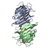
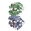
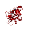
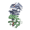
| Title | Crystal structure of human deoxycytidine kinase in complex with deoxycytidine | ||||||
 Components Components | Deoxycytidine kinase | ||||||
 Keywords Keywords | TRANSFERASE / Nucleoside kinase | ||||||
| Function / homology |  Function and homology informationdeoxycytidine kinase / 2'-deoxyadenosine kinase / deoxyguanosine kinase / dAMP salvage / deoxycytidine kinase activity / nucleoside phosphate biosynthetic process / deoxyguanosine kinase activity / deoxyadenosine kinase activity / Pyrimidine salvage / cytidine kinase activity ...deoxycytidine kinase / 2'-deoxyadenosine kinase / deoxyguanosine kinase / dAMP salvage / deoxycytidine kinase activity / nucleoside phosphate biosynthetic process / deoxyguanosine kinase activity / deoxyadenosine kinase activity / Pyrimidine salvage / cytidine kinase activity / pyrimidine nucleotide metabolic process / Purine salvage / phosphorylation / protein homodimerization activity / mitochondrion / nucleoplasm / ATP binding / cytosol / cytoplasm Function and homology informationdeoxycytidine kinase / 2'-deoxyadenosine kinase / deoxyguanosine kinase / dAMP salvage / deoxycytidine kinase activity / nucleoside phosphate biosynthetic process / deoxyguanosine kinase activity / deoxyadenosine kinase activity / Pyrimidine salvage / cytidine kinase activity ...deoxycytidine kinase / 2'-deoxyadenosine kinase / deoxyguanosine kinase / dAMP salvage / deoxycytidine kinase activity / nucleoside phosphate biosynthetic process / deoxyguanosine kinase activity / deoxyadenosine kinase activity / Pyrimidine salvage / cytidine kinase activity / pyrimidine nucleotide metabolic process / Purine salvage / phosphorylation / protein homodimerization activity / mitochondrion / nucleoplasm / ATP binding / cytosol / cytoplasmSimilarity search - Function | ||||||
| Biological species |  Homo sapiens (human) Homo sapiens (human) | ||||||
| Method | X-RAY DIFFRACTION / SYNCHROTRON / MOLECULAR REPLACEMENT / Resolution: 3.02 Å | ||||||
 Authors Authors | Godsey, M.H. / Ort, S. / Sabini, E. / Konrad, M. / Lavie, A. | ||||||
 Citation Citation | Journal: Biochemistry / Year: 2006 Title: Structural basis for the preference of UTP over ATP in human deoxycytidine kinase: illuminating the role of main-chain reorganization. Authors: Godsey, M.H. / Ort, S. / Sabini, E. / Konrad, M. / Lavie, A. | ||||||
| History |
| ||||||
| Remark 600 | HETEROGEN DCZ 302 are assoicated with protein chain A DCZ 402 are assoicated with protein chain B ...HETEROGEN DCZ 302 are assoicated with protein chain A DCZ 402 are assoicated with protein chain B DCZ 502 are assoicated with protein chain C DCZ 602 are assoicated with protein chain D |
- Structure visualization
| Structure viewer | Molecule: MolmilJmol/JSmol |
|---|
- Downloads & links
-Download
| PDBx/mmCIF format | 2a30.cif.gz | 187.4 KB | Display | PDBx/mmCIF format |
|---|---|---|---|---|
| PDB format | pdb2a30.ent.gz | 146.1 KB | Display | PDB format |
| PDBx/mmJSON format | 2a30.json.gz | Tree view | PDBx/mmJSON format | |
| Others |  Other downloads Other downloads |
-Validation report
| Arichive directory | https://data.pdbj.org/pub/pdb/validation_reports/a3/2a30ftp://data.pdbj.org/pub/pdb/validation_reports/a3/2a30 | HTTPS FTP |
|---|
-Related structure data
| Related structure data |  2a2zC  1p60S C: citing same article ( S: Starting model for refinement |
|---|---|
| Similar structure data |


-Links
 PDBj
PDBj
- Assembly
| Deposited unit | 
| ||||||||
|---|---|---|---|---|---|---|---|---|---|
| 1 | 
| ||||||||
| 2 | 
| ||||||||
| Unit cell |
| ||||||||
| Details | Biological assembly is a dimer. There are two dimers per ASU. |
-Components
| #1: Protein | / dCK Mass: 29139.004 Da / Num. of mol.: 4 / Mutation: residues 65-79 deletion Source method: isolated from a genetically manipulated source Source: (gene. exp.) Homo sapiens (human) / Gene: DCK / Plasmid: pET14b / Species (production host): Escherichia coli / Production host:  Escherichia coli BL21(DE3) (bacteria) / Strain (production host): BL21DE3 / References: UniProt: P27707, deoxycytidine kinase Escherichia coli BL21(DE3) (bacteria) / Strain (production host): BL21DE3 / References: UniProt: P27707, deoxycytidine kinase#2: Chemical |   Mass: 40.078 Da / Num. of mol.: 2 / Source method: obtained synthetically / Formula: Ca Mass: 40.078 Da / Num. of mol.: 2 / Source method: obtained synthetically / Formula: Ca#3: Chemical | ChemComp-DCZ / Deoxycytidine  Type: DNA OH 5 prime terminus / Mass: 227.217 Da / Num. of mol.: 4 / Source method: obtained synthetically / Formula: C9H13N3O4 Type: DNA OH 5 prime terminus / Mass: 227.217 Da / Num. of mol.: 4 / Source method: obtained synthetically / Formula: C9H13N3O4#4: Water | ChemComp-HOH / | Water Mass: 18.015 Da / Num. of mol.: 26 / Source method: isolated from a natural source / Formula: H2O Mass: 18.015 Da / Num. of mol.: 26 / Source method: isolated from a natural source / Formula: H2O |
|---|
-Experimental details
-Experiment
| Experiment | Method: X-RAY DIFFRACTION / Number of used crystals: 1 |
|---|
- Sample preparation
| Crystal | Density Matthews: 2.4 Å3/Da / Density % sol: 48.1 % |
|---|---|
| Crystal grow | Temperature: 298 K / Method: vapor diffusion, hanging drop / pH: 6.5 Details: PEG 1000, MPD, Calcium Chloride, Sodium Cacodylate, pH 6.5, VAPOR DIFFUSION, HANGING DROP, temperature 298K |
-Data collection
| Diffraction | Mean temperature: 100 K |
|---|---|
| Diffraction source | Source: SYNCHROTRON / Site: APS  / Beamline: 22-ID / Wavelength: 0.97626 Å / Beamline: 22-ID / Wavelength: 0.97626 Å |
| Detector | Type: MARRESEARCH / Detector: CCD / Date: Apr 8, 2004 / Details: MIRROR |
| Radiation | Monochromator: Sagittaly focused Si (220) / Protocol: SINGLE WAVELENGTH / Monochromatic (M) / Laue (L): M / Scattering type: x-ray |
| Radiation wavelength | Wavelength: 0.97626 Å / Relative weight: 1 |
| Reflection | Resolution: 3→40 Å / Num. all: 22365 / Num. obs: 20040 / % possible obs: 83.7 % / Observed criterion σ(F): 0 / Observed criterion σ(I): 0 / Redundancy: 3.3 % / Rmerge(I) obs: 0.141 / Rsym value: 0.118 / Net I/σ(I): 8.77 |
| Reflection shell | Resolution: 3→3.1 Å / % possible obs: 53.4 % / Redundancy: 3.2 % / Rmerge(I) obs: 0.642 / Mean I/σ(I) obs: 2.36 / Num. measured obs: 3602 / Num. unique all: 1124 / Num. unique obs: 1124 / Rsym value: 0.542 / % possible all: 81.2 |
-Phasing
| Phasing MR | Rfactor: 52.1 / Cor.coef. Fo:Fc: 37.2 / Cor.coef. Io to Ic: 42.9
|
|---|
- Processing
| Software |
| ||||||||||||||||||||||||||||||||||||||||||||||||||||||||||||||||||||||||||||||||||||||||||
|---|---|---|---|---|---|---|---|---|---|---|---|---|---|---|---|---|---|---|---|---|---|---|---|---|---|---|---|---|---|---|---|---|---|---|---|---|---|---|---|---|---|---|---|---|---|---|---|---|---|---|---|---|---|---|---|---|---|---|---|---|---|---|---|---|---|---|---|---|---|---|---|---|---|---|---|---|---|---|---|---|---|---|---|---|---|---|---|---|---|---|---|
| Refinement | Method to determine structure: MOLECULAR REPLACEMENT Starting model: PDB entry 1P60 Resolution: 3.02→19.9 Å / Rfactor Rfree error: 0.006 / Occupancy max: 1 / Occupancy min: 1 / Cross valid method: THROUGHOUT / σ(F): 0 / σ(I): 0 / Stereochemistry target values: Engh & Huber
| ||||||||||||||||||||||||||||||||||||||||||||||||||||||||||||||||||||||||||||||||||||||||||
| Solvent computation | Solvent model: CNS bulk solvent model used / Bsol: 34.1324 Å2 / ksol: 0.230619 e/Å3 | ||||||||||||||||||||||||||||||||||||||||||||||||||||||||||||||||||||||||||||||||||||||||||
| Displacement parameters | Biso max: 109.84 Å2 / Biso mean: 69.89 Å2 / Biso min: 22.85 Å2
| ||||||||||||||||||||||||||||||||||||||||||||||||||||||||||||||||||||||||||||||||||||||||||
| Refine analyze |
| ||||||||||||||||||||||||||||||||||||||||||||||||||||||||||||||||||||||||||||||||||||||||||
| Refinement step | Cycle: LAST / Resolution: 3.02→19.9 Å
| ||||||||||||||||||||||||||||||||||||||||||||||||||||||||||||||||||||||||||||||||||||||||||
| Refine LS restraints |
| ||||||||||||||||||||||||||||||||||||||||||||||||||||||||||||||||||||||||||||||||||||||||||
| LS refinement shell | Refine-ID: X-RAY DIFFRACTION
|
