 Movie
Movie Controller


+ Open data

- Basic information
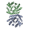
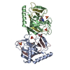
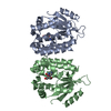
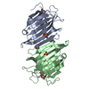
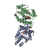
| Entry | Database: PDB / ID: 1itv | ||||||
|---|---|---|---|---|---|---|---|
| Title | Dimeric form of the haemopexin domain of MMP9 | ||||||
 Components Components | MMP9 | ||||||
 Keywords Keywords |  HYDROLASE / MMP9 / adaptive molecular recognition / beta propeller / dimer HYDROLASE / MMP9 / adaptive molecular recognition / beta propeller / dimer | ||||||
| Function / homology |  Function and homology informationgelatinase B / negative regulation of epithelial cell differentiation involved in kidney development / negative regulation of cation channel activity / negative regulation of cysteine-type endopeptidase activity involved in apoptotic signaling pathway / cellular response to UV-A / positive regulation of keratinocyte migration / regulation of neuroinflammatory response / Assembly of collagen fibrils and other multimeric structures / positive regulation of epidermal growth factor receptor signaling pathway / Activation of Matrix Metalloproteinases ...gelatinase B / negative regulation of epithelial cell differentiation involved in kidney development / negative regulation of cation channel activity / negative regulation of cysteine-type endopeptidase activity involved in apoptotic signaling pathway / cellular response to UV-A / positive regulation of keratinocyte migration / regulation of neuroinflammatory response / Assembly of collagen fibrils and other multimeric structures / positive regulation of epidermal growth factor receptor signaling pathway / Activation of Matrix Metalloproteinases / endodermal cell differentiation / positive regulation of release of cytochrome c from mitochondria / response to amyloid-beta / macrophage differentiation / Collagen degradation / EPH-ephrin mediated repulsion of cells / collagen catabolic process / extracellular matrix disassembly / ephrin receptor signaling pathway / positive regulation of DNA binding / negative regulation of intrinsic apoptotic signaling pathway / embryo implantation / positive regulation of vascular associated smooth muscle cell proliferation / cellular response to cadmium ion / collagen binding / Degradation of the extracellular matrix / extracellular matrix organization / positive regulation of receptor binding / skeletal system development / Signaling by SCF-KIT / metalloendopeptidase activity / cellular response to reactive oxygen species / metallopeptidase activity / cell migration / tertiary granule lumen / peptidase activity / collagen-containing extracellular matrix / Interleukin-4 and Interleukin-13 signaling / endopeptidase activity / cellular response to lipopolysaccharide / ficolin-1-rich granule lumen / Extra-nuclear estrogen signaling / positive regulation of protein phosphorylation / positive regulation of apoptotic process / serine-type endopeptidase activity / apoptotic process / Neutrophil degranulation / negative regulation of apoptotic process / proteolysis / extracellular space / extracellular exosome / zinc ion binding / extracellular region / identical protein binding Function and homology informationgelatinase B / negative regulation of epithelial cell differentiation involved in kidney development / negative regulation of cation channel activity / negative regulation of cysteine-type endopeptidase activity involved in apoptotic signaling pathway / cellular response to UV-A / positive regulation of keratinocyte migration / regulation of neuroinflammatory response / Assembly of collagen fibrils and other multimeric structures / positive regulation of epidermal growth factor receptor signaling pathway / Activation of Matrix Metalloproteinases ...gelatinase B / negative regulation of epithelial cell differentiation involved in kidney development / negative regulation of cation channel activity / negative regulation of cysteine-type endopeptidase activity involved in apoptotic signaling pathway / cellular response to UV-A / positive regulation of keratinocyte migration / regulation of neuroinflammatory response / Assembly of collagen fibrils and other multimeric structures / positive regulation of epidermal growth factor receptor signaling pathway / Activation of Matrix Metalloproteinases / endodermal cell differentiation / positive regulation of release of cytochrome c from mitochondria / response to amyloid-beta / macrophage differentiation / Collagen degradation / EPH-ephrin mediated repulsion of cells / collagen catabolic process / extracellular matrix disassembly / ephrin receptor signaling pathway / positive regulation of DNA binding / negative regulation of intrinsic apoptotic signaling pathway / embryo implantation / positive regulation of vascular associated smooth muscle cell proliferation / cellular response to cadmium ion / collagen binding / Degradation of the extracellular matrix / extracellular matrix organization / positive regulation of receptor binding / skeletal system development / Signaling by SCF-KIT / metalloendopeptidase activity / cellular response to reactive oxygen species / metallopeptidase activity / cell migration / tertiary granule lumen / peptidase activity / collagen-containing extracellular matrix / Interleukin-4 and Interleukin-13 signaling / endopeptidase activity / cellular response to lipopolysaccharide / ficolin-1-rich granule lumen / Extra-nuclear estrogen signaling / positive regulation of protein phosphorylation / positive regulation of apoptotic process / serine-type endopeptidase activity / apoptotic process / Neutrophil degranulation / negative regulation of apoptotic process / proteolysis / extracellular space / extracellular exosome / zinc ion binding / extracellular region / identical protein bindingSimilarity search - Function | ||||||
| Biological species |  Homo sapiens (human) Homo sapiens (human) | ||||||
| Method | X-RAY DIFFRACTION / SYNCHROTRON / MIR / Resolution: 1.95 Å | ||||||
 Authors Authors | Cha, H. / Kopetzki, E. / Huber, R. / Lanzendoerfer, M. / Brandstetter, H. | ||||||
 Citation Citation | Journal: J.Mol.Biol. / Year: 2002 Title: Structural basis of the adaptive molecular recognition by MMP9. Authors: Cha, H. / Kopetzki, E. / Huber, R. / Lanzendorfer, M. / Brandstetter, H. | ||||||
| History |
|
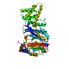
- Structure visualization
| Structure viewer | Molecule: MolmilJmol/JSmol |
|---|
- Downloads & links
-Download
| PDBx/mmCIF format | 1itv.cif.gz | 94 KB | Display | PDBx/mmCIF format |
|---|---|---|---|---|
| PDB format | pdb1itv.ent.gz | 71.8 KB | Display | PDB format |
| PDBx/mmJSON format | 1itv.json.gz | Tree view | PDBx/mmJSON format | |
| Others |  Other downloads Other downloads |
-Validation report
| Arichive directory | https://data.pdbj.org/pub/pdb/validation_reports/it/1itvftp://data.pdbj.org/pub/pdb/validation_reports/it/1itv | HTTPS FTP |
|---|
-Related structure data
| Similar structure data |
|---|


-Links
 PDBj
PDBj


- Assembly
| Deposited unit | 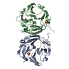
| ||||||||
|---|---|---|---|---|---|---|---|---|---|
| 1 |
| ||||||||
| Unit cell |
| ||||||||
| Details | the asu forms the biological dimer |
-Components
| #1: Protein | Mass: 22450.525 Da / Num. of mol.: 2 / Fragment: haemopexin-like domain Source method: isolated from a genetically manipulated source Source: (gene. exp.) Homo sapiens (human) / Production host:  Escherichia coli (E. coli) / References: UniProt: P14780, gelatinase B Escherichia coli (E. coli) / References: UniProt: P14780, gelatinase B#2: Chemical | ChemComp-SO4 / Sulfate  Mass: 96.063 Da / Num. of mol.: 4 / Source method: obtained synthetically / Formula: SO4 Mass: 96.063 Da / Num. of mol.: 4 / Source method: obtained synthetically / Formula: SO4#3: Water | ChemComp-HOH / | Water Mass: 18.015 Da / Num. of mol.: 182 / Source method: isolated from a natural source / Formula: H2O Mass: 18.015 Da / Num. of mol.: 182 / Source method: isolated from a natural source / Formula: H2O |
|---|
-Experimental details
-Experiment
| Experiment | Method: X-RAY DIFFRACTION / Number of used crystals: 1 |
|---|
- Sample preparation
| Crystal | Density Matthews: 2.85 Å3/Da / Density % sol: 56.82 % | ||||||||||||||||||||||||||||||
|---|---|---|---|---|---|---|---|---|---|---|---|---|---|---|---|---|---|---|---|---|---|---|---|---|---|---|---|---|---|---|---|
| Crystal grow | Temperature: 291 K / Method: vapor diffusion, sitting drop / pH: 7.5 Details: Tris, PEGMME, ammonium sulphate, pH 7.5, VAPOR DIFFUSION, SITTING DROP, temperature 291K | ||||||||||||||||||||||||||||||
| Crystal grow | *PLUS | ||||||||||||||||||||||||||||||
| Components of the solutions | *PLUS
|
-Data collection
| Diffraction |
| |||||||||
|---|---|---|---|---|---|---|---|---|---|---|
| Diffraction source | Source: SYNCHROTRON / Site: MPG/DESY, HAMBURG  / Beamline: BW6 / Wavelength: 1.05 Å / Beamline: BW6 / Wavelength: 1.05 Å | |||||||||
| Detector | Type: MARRESEARCH / Detector: CCD / Date: Mar 3, 2001 | |||||||||
| Radiation | Protocol: MAD / Monochromatic (M) / Laue (L): M / Scattering type: x-ray | |||||||||
| Radiation wavelength | Wavelength: 1.05 Å / Relative weight: 1 | |||||||||
| Reflection | Resolution: 1.95→20 Å / Num. all: 213889 / Num. obs: 36949 / % possible obs: 97.8 % / Observed criterion σ(F): 2 / Observed criterion σ(I): 2 / Rmerge(I) obs: 0.082 | |||||||||
| Reflection shell | Resolution: 1.95→20 Å / % possible all: 97.8 | |||||||||
| Reflection | *PLUS Lowest resolution: 20 Å / Num. measured all: 124981 |
- Processing
| Software |
| ||||||||||||||||||||
|---|---|---|---|---|---|---|---|---|---|---|---|---|---|---|---|---|---|---|---|---|---|
| Refinement | Method to determine structure: MIR / Resolution: 1.95→20 Å / σ(F): 2 / Stereochemistry target values: Engh & Huber
| ||||||||||||||||||||
| Refinement step | Cycle: LAST / Resolution: 1.95→20 Å
| ||||||||||||||||||||
| Refine LS restraints |
|
