 Movie
Movie Controller

[English] 日本語
 Yorodumi
- PDB-1xmk: The Crystal structure of the Zb domain from the RNA editing enzym... -
+ Open data

- Basic information
| Entry | Database: PDB / ID: 1xmk | ||||||
|---|---|---|---|---|---|---|---|
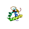
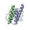
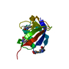
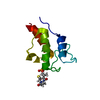
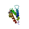
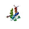
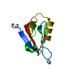
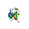
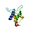
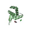
| Title | The Crystal structure of the Zb domain from the RNA editing enzyme ADAR1 | ||||||
 Components Components | Double-stranded RNA-specific adenosine deaminase | ||||||
 Keywords Keywords |  HYDROLASE / winged Helix-Turn-Helix / RNA editing / Interferon / ADAR1 HYDROLASE / winged Helix-Turn-Helix / RNA editing / Interferon / ADAR1 | ||||||
| Function / homology |  Function and homology information Function and homology informationsomatic diversification of immune receptors via somatic mutation / negative regulation of post-transcriptional gene silencing by regulatory ncRNA / C6 deamination of adenosine / Formation of editosomes by ADAR proteins / double-stranded RNA adenine deaminase / tRNA-specific adenosine deaminase activity / supraspliceosomal complex / double-stranded RNA adenosine deaminase activity / negative regulation of protein kinase activity by regulation of protein phosphorylation / base conversion or substitution editing ...somatic diversification of immune receptors via somatic mutation / negative regulation of post-transcriptional gene silencing by regulatory ncRNA / C6 deamination of adenosine / Formation of editosomes by ADAR proteins / double-stranded RNA adenine deaminase / tRNA-specific adenosine deaminase activity / supraspliceosomal complex / double-stranded RNA adenosine deaminase activity / negative regulation of protein kinase activity by regulation of protein phosphorylation / base conversion or substitution editing / hematopoietic stem cell homeostasis / response to interferon-alpha / adenosine to inosine editing / RISC complex assembly / pre-miRNA processing / negative regulation of hepatocyte apoptotic process / definitive hemopoiesis / negative regulation of type I interferon-mediated signaling pathway / hepatocyte apoptotic process / positive regulation of viral genome replication / RNA processing / hematopoietic progenitor cell differentiation / protein export from nucleus / erythrocyte differentiation / response to virus / PKR-mediated signaling / mRNA processing / cellular response to virus / osteoblast differentiation / protein import into nucleus / double-stranded RNA binding / Interferon alpha/beta signaling / defense response to virus / innate immune response / nucleolus / DNA binding / RNA binding / nucleoplasm / membrane / metal ion binding / nucleus / cytosol / cytoplasmSimilarity search - Function | ||||||
| Biological species |  Homo sapiens (human) Homo sapiens (human) | ||||||
| Method | X-RAY DIFFRACTION / SYNCHROTRON / MIR / Resolution: 0.97 Å | ||||||
 Authors Authors | Athanasiadis, A. / Placido, D. / Maas, S. / Brown II, B.A. / Lowenhaupt, K. / Rich, A. | ||||||
 Citation Citation | Journal: J.Mol.Biol. / Year: 2005 Title: The Crystal Structure of the Z[beta] Domain of the RNA-editing Enzyme ADAR1 Reveals Distinct Conserved Surfaces Among Z-domains. Authors: Athanasiadis, A. / Placido, D. / Maas, S. / Brown II, B.A. / Lowenhaupt, K. / Rich, A. #1: Journal: Nat.Struct.Mol.Biol. / Year: 2001Title: Structure of the DLM-1-Z-DNA complex reveals a conserved family of Z-DNA-binding proteins Authors: Schwartz, T. / Behlke, J. / Lowenhaupt, K. / Heinemann, U. / Rich, A. #2: Journal: Science / Year: 1999Title: Crystal structure of the Zalpha domain of the human editing enzyme ADAR1 bound to left-handed Z-DNA Authors: Schwartz, T. / Rould, M.A. / Lowenhaupt, K. / Herbert, A. / Rich, A. | ||||||
| History |
|
- Structure visualization
| Structure viewer | Molecule: MolmilJmol/JSmol |
|---|
- Downloads & links
-Download
| PDBx/mmCIF format | 1xmk.cif.gz | 54 KB | Display | PDBx/mmCIF format |
|---|---|---|---|---|
| PDB format | pdb1xmk.ent.gz | 37.8 KB | Display | PDB format |
| PDBx/mmJSON format | 1xmk.json.gz | Tree view | PDBx/mmJSON format | |
| Others |  Other downloads Other downloads |
-Validation report
| Arichive directory | https://data.pdbj.org/pub/pdb/validation_reports/xm/1xmkftp://data.pdbj.org/pub/pdb/validation_reports/xm/1xmk | HTTPS FTP |
|---|
-Related structure data
| Related structure data | |
|---|---|
| Similar structure data |


-Links
 PDBj
PDBj

- Assembly
| Deposited unit | 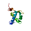
| ||||||||
|---|---|---|---|---|---|---|---|---|---|
| 1 |
| ||||||||
| Unit cell |
|
-Components
| #1: Protein | Mass: 9005.369 Da / Num. of mol.: 1 Source method: isolated from a genetically manipulated source Source: (gene. exp.) Homo sapiens (human) / Gene: ADAR, ADAR1, DSRAD, IFI4 / Plasmid: pET28 / Species (production host): Escherichia coli / Production host:  Escherichia coli BL21(DE3) (bacteria) / Strain (production host): BL21(DE3) Escherichia coli BL21(DE3) (bacteria) / Strain (production host): BL21(DE3)References: UniProt: P55265, Hydrolases; Acting on carbon-nitrogen bonds, other than peptide bonds; In cyclic amidines | ||||||
|---|---|---|---|---|---|---|---|
| #2: Chemical |   Mass: 112.411 Da / Num. of mol.: 2 / Source method: obtained synthetically / Formula: Cd Mass: 112.411 Da / Num. of mol.: 2 / Source method: obtained synthetically / Formula: Cd#3: Chemical | ChemComp-NI / | Nickel  Mass: 58.693 Da / Num. of mol.: 1 / Source method: obtained synthetically / Formula: Ni Mass: 58.693 Da / Num. of mol.: 1 / Source method: obtained synthetically / Formula: Ni#4: Chemical | Chloride  Mass: 35.453 Da / Num. of mol.: 2 / Source method: obtained synthetically / Formula: Cl Mass: 35.453 Da / Num. of mol.: 2 / Source method: obtained synthetically / Formula: Cl#5: Water | ChemComp-HOH / | Water Mass: 18.015 Da / Num. of mol.: 125 / Source method: isolated from a natural source / Formula: H2O Mass: 18.015 Da / Num. of mol.: 125 / Source method: isolated from a natural source / Formula: H2O |
-Experimental details
-Experiment
| Experiment | Method: X-RAY DIFFRACTION / Number of used crystals: 1 |
|---|
- Sample preparation
| Crystal | Density Matthews: 1.7 Å3/Da / Density % sol: 27.5 % |
|---|---|
| Crystal grow | Temperature: 312 K / pH: 9 Details: PEG1000, Cadmium Chloride, Nickel Chloride, Tris, pH 9.0, VAPOR DIFFUSION, HANGING DROP, temperature 312K, pH 9.00 |
-Data collection
| Diffraction | Mean temperature: 100 K |
|---|---|
| Diffraction source | Source: SYNCHROTRON / Site: NSLS  / Beamline: X8C / Wavelength: 0.9 / Beamline: X8C / Wavelength: 0.9 |
| Detector | Type: ADSC QUANTUM 4 / Detector: CCD / Date: Jan 22, 2002 |
| Radiation | Monochromator: MONOCHROMATOR / Protocol: SINGLE WAVELENGTH / Monochromatic (M) / Laue (L): M / Scattering type: x-ray |
| Radiation wavelength | Wavelength: 0.9 Å / Relative weight: 1 |
| Reflection | Resolution: 0.97→27.92 Å / Num. obs: 41638 / % possible obs: 99.2 % / Observed criterion σ(I): 1 / Redundancy: 4.1 % / Biso Wilson estimate: 6.5 Å2 / Rsym value: 0.075 / Net I/σ(I): 5.2 |
| Reflection shell | Resolution: 0.97→1.02 Å / % possible all: 94.7 |
- Processing
| Software |
| |||||||||||||||||||||||||||||||||
|---|---|---|---|---|---|---|---|---|---|---|---|---|---|---|---|---|---|---|---|---|---|---|---|---|---|---|---|---|---|---|---|---|---|---|
| Refinement | Method to determine structure: MIR / Resolution: 0.97→10 Å Cross valid method: THROUGHOUT WITH THE EXCEPTION OF THE LAST TWO REFINEMENT CYCLES σ(F): 0 / Stereochemistry target values: Engh & Huber / Details: CNS WAS USED IN EARLY REFINEMENT stages
| |||||||||||||||||||||||||||||||||
| Solvent computation | Solvent model: MOEWS & KRETSINGER, J.mol.biol.91(1973)201-228 | |||||||||||||||||||||||||||||||||
| Refine analyze | Num. disordered residues: 4 | |||||||||||||||||||||||||||||||||
| Refinement step | Cycle: LAST / Resolution: 0.97→10 Å
| |||||||||||||||||||||||||||||||||
| Refine LS restraints |
|
