 Movie
Movie Controller

[English] 日本語
 Yorodumi
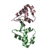
- PDB-2fu4: Crystal Structure of the DNA binding domain of E.coli FUR (Ferric... -
+ Open data

- Basic information
| Entry | Database: PDB / ID: 2fu4 | ||||||
|---|---|---|---|---|---|---|---|
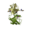
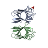
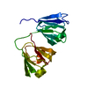
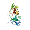
| Title | Crystal Structure of the DNA binding domain of E.coli FUR (Ferric Uptake Regulator) | ||||||
 Components Components | Ferric uptake regulation protein | ||||||
 Keywords Keywords |  DNA BINDING PROTEIN / DNA binding domain / helix-turn-helix DNA BINDING PROTEIN / DNA binding domain / helix-turn-helix | ||||||
| Function / homology |  Function and homology information Function and homology informationnegative regulation of siderophore biosynthetic process / DNA-binding transcription repressor activity / DNA-binding transcription activator activity / protein-DNA complex / sequence-specific DNA binding / transcription cis-regulatory region binding / DNA-binding transcription factor activity / negative regulation of DNA-templated transcription / positive regulation of DNA-templated transcription / zinc ion binding / cytosolSimilarity search - Function | ||||||
| Biological species |  Escherichia coli (E. coli) Escherichia coli (E. coli) | ||||||
| Method | X-RAY DIFFRACTION / SYNCHROTRON / MOLECULAR REPLACEMENT / Resolution: 1.8 Å | ||||||
 Authors Authors | Pecqueur, L. / D'Autreaux, B. / Dupuy, J. / Nicolet, Y. / Jacquamet, L. / Brutscher, B. / Michaud-Soret, I. / Bersch, B. | ||||||
 Citation Citation | Journal: J.Biol.Chem. / Year: 2006 Title: Structural changes of Escherichia coli ferric uptake regulator during metal-dependent dimerization and activation explored by NMR and X-ray crystallography Authors: Pecqueur, L. / D'Autreaux, B. / Dupuy, J. / Nicolet, Y. / Jacquamet, L. / Brutscher, B. / Michaud-Soret, I. / Bersch, B. | ||||||
| History |
|
- Structure visualization
| Structure viewer | Molecule: MolmilJmol/JSmol |
|---|
- Downloads & links
-Download
| PDBx/mmCIF format | 2fu4.cif.gz | 84.4 KB | Display | PDBx/mmCIF format |
|---|---|---|---|---|
| PDB format | pdb2fu4.ent.gz | 64.7 KB | Display | PDB format |
| PDBx/mmJSON format | 2fu4.json.gz | Tree view | PDBx/mmJSON format | |
| Others |  Other downloads Other downloads |
-Validation report
| Arichive directory | https://data.pdbj.org/pub/pdb/validation_reports/fu/2fu4ftp://data.pdbj.org/pub/pdb/validation_reports/fu/2fu4 | HTTPS FTP |
|---|
-Related structure data
| Similar structure data |
|---|


-Links
 PDBj
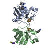
PDBj- Assembly
| Deposited unit | 
| ||||||||
|---|---|---|---|---|---|---|---|---|---|
| 1 |
| ||||||||
| 2 |
| ||||||||
| 3 |
| ||||||||
| 4 | 
| ||||||||
| 5 | 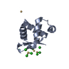
| ||||||||
| 6 | 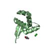
| ||||||||
| Unit cell |
|
-Components
| #1: Protein | Mass: 9351.656 Da / Num. of mol.: 2 / Fragment: N-terminal domain Source method: isolated from a genetically manipulated source Source: (gene. exp.) Escherichia coli (E. coli) / Strain: BL21List of strains of Escherichia coli / Gene: FUR(P0A9A9) / Plasmid: pET 30c / Production host: Escherichia coli (E. coli) / Strain (production host): BL21 / References: UniProt: P0A9A9#2: Chemical | ChemComp-CD /   Mass: 112.411 Da / Num. of mol.: 8 / Source method: obtained synthetically / Formula: Cd Mass: 112.411 Da / Num. of mol.: 8 / Source method: obtained synthetically / Formula: Cd#3: Chemical | ChemComp-CL / Chloride  Mass: 35.453 Da / Num. of mol.: 12 / Source method: obtained synthetically / Formula: Cl Mass: 35.453 Da / Num. of mol.: 12 / Source method: obtained synthetically / Formula: Cl#4: Chemical | ChemComp-GOL / | Glycerol  Mass: 92.094 Da / Num. of mol.: 1 / Source method: obtained synthetically / Formula: C3H8O3 Mass: 92.094 Da / Num. of mol.: 1 / Source method: obtained synthetically / Formula: C3H8O3#5: Water | ChemComp-HOH / | Water Mass: 18.015 Da / Num. of mol.: 160 / Source method: isolated from a natural source / Formula: H2O Mass: 18.015 Da / Num. of mol.: 160 / Source method: isolated from a natural source / Formula: H2O |
|---|
-Experimental details
-Experiment
| Experiment | Method: X-RAY DIFFRACTION / Number of used crystals: 1 |
|---|
- Sample preparation
| Crystal | Density Matthews: 2.35 Å3/Da / Density % sol: 47.61 % |
|---|---|
| Crystal grow | Temperature: 293 K / Method: vapor diffusion, hanging drop / pH: 4.6 Details: 100mM sodium acetate, 30%(v/v) PEG 200 containing 100mM CdCl2, pH 4.6, VAPOR DIFFUSION, HANGING DROP, temperature 293K |
-Data collection
| Diffraction | Mean temperature: 100 K |
|---|---|
| Diffraction source | Source: SYNCHROTRON / Site: ESRF  / Beamline: BM30A / Wavelength: 0.979637 Å / Beamline: BM30A / Wavelength: 0.979637 Å |
| Detector | Type: MARRESEARCH / Detector: CCD / Date: Sep 7, 2005 |
| Radiation | Monochromator: Si 111 / Protocol: SINGLE WAVELENGTH / Monochromatic (M) / Laue (L): M / Scattering type: x-ray |
| Radiation wavelength | Wavelength: 0.979637 Å / Relative weight: 1 |
| Reflection | Resolution: 1.8→50 Å / Num. obs: 17195 / % possible obs: 99.4 % / Observed criterion σ(F): 2 / Observed criterion σ(I): 2 / Redundancy: 8.5 % / Biso Wilson estimate: 16.6 Å2 / Rsym value: 0.092 / Net I/σ(I): 17.72 |
| Reflection shell | Resolution: 1.8→1.9 Å / Redundancy: 8.4 % / Mean I/σ(I) obs: 6.2 / Num. unique all: 22370 / Rsym value: 0.329 / % possible all: 97.1 |
- Processing
| Software |
| |||||||||||||||||||||||||||||||||||||||||||||||||||||||||||||||||||||||||||||||||||||||||||||||||||||||||||||||||||
|---|---|---|---|---|---|---|---|---|---|---|---|---|---|---|---|---|---|---|---|---|---|---|---|---|---|---|---|---|---|---|---|---|---|---|---|---|---|---|---|---|---|---|---|---|---|---|---|---|---|---|---|---|---|---|---|---|---|---|---|---|---|---|---|---|---|---|---|---|---|---|---|---|---|---|---|---|---|---|---|---|---|---|---|---|---|---|---|---|---|---|---|---|---|---|---|---|---|---|---|---|---|---|---|---|---|---|---|---|---|---|---|---|---|---|---|---|
| Refinement | Method to determine structure: MOLECULAR REPLACEMENT / Resolution: 1.8→37.42 Å / Cor.coef. Fo:Fc: 0.941 / Cor.coef. Fo:Fc free: 0.913 / SU B: 5.446 / SU ML: 0.078 / Cross valid method: THROUGHOUT / σ(F): 2 / σ(I): 2 / ESU R: 0.225 / ESU R Free: 0.119 / Stereochemistry target values: MAXIMUM LIKELIHOOD
| |||||||||||||||||||||||||||||||||||||||||||||||||||||||||||||||||||||||||||||||||||||||||||||||||||||||||||||||||||
| Solvent computation | Ion probe radii: 0.8 Å / Shrinkage radii: 0.8 Å / VDW probe radii: 1.2 Å / Solvent model: MASK | |||||||||||||||||||||||||||||||||||||||||||||||||||||||||||||||||||||||||||||||||||||||||||||||||||||||||||||||||||
| Displacement parameters | Biso mean: 16.613 Å2 | |||||||||||||||||||||||||||||||||||||||||||||||||||||||||||||||||||||||||||||||||||||||||||||||||||||||||||||||||||
| Refinement step | Cycle: LAST / Resolution: 1.8→37.42 Å
| |||||||||||||||||||||||||||||||||||||||||||||||||||||||||||||||||||||||||||||||||||||||||||||||||||||||||||||||||||
| Refine LS restraints |
| |||||||||||||||||||||||||||||||||||||||||||||||||||||||||||||||||||||||||||||||||||||||||||||||||||||||||||||||||||
| LS refinement shell | Resolution: 1.795→1.842 Å / Total num. of bins used: 20
|
